Tongue Base Schwannoma: A Case Report and Literature Review
Download
Abstract
Background: Schwannoma is a benign tumor arises from Schwann cells of the peripheral nerves. Tongue base schwannomas are very rare and have been sporadically reported; it is usually missed when evaluating a tongue base mass. This work aimed to report a new case of tongue base schwannoma and to review the literature about this tumor.
Case presentation: A male patient of 40 years old presented with slowly enlarging mass at the tongue base. Clinical and radiological findings highly suspected of tongue base benign lesion. Trans-oral resection was done and the specimen was subjected to the histopathological evaluation.
Conclusion: Schwannoma should be considered for a well-defined, painless, firm swelling at the tongue base.
Introduction
Schwannomas, also known as neurilemmomas, are encapsulated benign nerve sheath tumors. Schwanommas are usually solitary, slowly growing, and painless. It results from Schwann cell proliferation in the peripheral nerves [1]. Schwanomma is usually skipped out when evaluating a tongue base mass because it is not usually considered in the differential diagnosis of benign lesions affecting the tongue [2]. The diagnosis may be suspected clinically and radiologically, but it depends mainly on the histopathological confirmation [3]. In this article, we reported a new case and reviewed the literature about this tumor particularly in tongue base region.
Materials and Methods
This work conducted according to the local institutional ethical instructions. A written informed consent was obtained from the participant patient. Comprehensive review was collected using the keywords “Schwannoma; Neurilemmoma; Tongue base” via Medline/ Pubmed research. We selected only full English articles with confirmed diagnosis as schwannoma. Data were analyzed as regard the patients’ clinical characteristics including the sex, age, clinical presentation, tumor size and surgical approaches.
Case report
A 40 years old male presented with slowly growing swelling at the tongue base of unknown duration. The patient had no difficulty in breathing or swallowing. No history of pain, local bleeding and no impairment of the tongue movement. The patient was non-smoker.
On examination, a well-defined mass measuring about 2x2 cm was detected at the tongue base with normal mobility of the tongue and soft palate. On palpation, it was firm, non-tender, with no bleeding on touch, no overlying ulceration and it was not attached to surrounding tissues. MRI revealed well-defined oval soft tissue lesion at the tongue base with no infiltration of surrounding tissues. Trans-oral excision was done at the Department of General Surgery of Sohag University Hospital and the specimen was sent to the Pathology Laboratory of the same hospital.
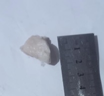
Gross examination showed well defined mass with slightly irregular surface, firm consistency, grayish white cut surface (Figure 1).
Figure 1. The Surgical Specimen Presented as Well-defined, Encapsulated Firm White Mass at the Base of Tongue.

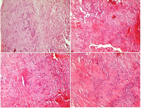
Histopathological examination of serial sections from the submitted specimen revealed benign neoplastic lesion formed of proliferating spindle-shaped cells arranged in short bundles. Focal hypocellular areas were seen. The cells have moderate pale eosinophilic cytoplasm and uniform nuclei. Frequent foci of nuclear palisading were seen. Histopathological features were consistent with the diagnosis of schwannoma (Figure 2).
Figure 2. Histologic Examination Revealed a Benign Neoplastic Lesion Composed of Spindle Shaped Cells Arranged in Short Bundles. Focal hypocellular areas were seen. The cells have moderate pale eosinophilic cytoplasm and short nuclei arranged in a characteristic palisading fashion with organoid arrangement (Verocay bodies). (H&E stain).

Discussion
Schwannomas are benign neurogenic tumors, firstly was described by Verocay. They arise from Schwann cells that comprise the myelin sheaths surrounding peripheral nerve. about 25-40% of schwannomas occur in the head and neck, of these, about 1-2% only affect the intraoral area. It is commonly seen at the tongue mainly, about 66%, in anterior third of the tongue [4].
Intraoral schwannomas affects any age, but commonly occur between 2nd and 4th decades of life. The reports about the sex predilection were controversy; Some reported that there was no sex predilection [5], while others reported male predominance [6]. Schwannomas are usually solitary, however multiple lesions may also be seen in neurofibromatosis type 1 or type 2 [7].
The most frequent clinical presentations are progressive dysphagia, globus sensation and painless swelling. Other complaints include odynophagia, pain, breathing difficulty, snoring and change of voice or asymptomatic were reported nearly in one fourth of patien. Lesion size was ranging from 1.2 to 7.9 cm. The commonest therapeutic approach was the Trans-oral approach [8].
Malignant transformation of a solitary extracranial head and neck schwannoma is extremely rare and it has been reported in one case in the tongue [9]. It is very difficult to distinguish schwannomas from other intra-oral well-defined encapsulated swellings clinically [10].
Histopathologically, schwannomas are classified into several subtypes including: classical, cellular, epitheloid, plexiform, melanotic, ancient, and granular cell schwannoma [11]. The characteristic histological picture of schwannomas is biphasic pattern formed of alternating hypercelullar Antoni A spindle cell areas and myxoid hypocellular Antoni B areas. Nuclear palisading around fibrillary process forms Verocay bodies. Hemorrhage, necrosis and mitotic activity are rarely encountered [12]. Immunohistochemical markers help to confirm the diagnosis and support the Schwann cell origin. Immunohistochemistry shows that schwannoma cells tongue exhibits strong and diffuse nuclear and cytoplasmic S-100 protein expression and extensive nuclear SOX10 [13].
Radiologically, MRI is superior to CT for schwannomas allows a more precise localization and better visualization of the relations to other structures, as well as a more accurate measurement of tumor size. Moreover, these tumors usually appear smooth, well demarcated, and do not invade the surrounding musculature [5].
In conclusion, Tongue base schwannomas should be considered in differential diagnosis of tongue mass. The diagnosis should be confirmed after histopathological examination and supported by immunohistochemical analysis in challenging cases.
Conflict of Interest
The authors declare that they have no competing interests.
References
- Clinical Features and Surgical Treatment of Schwannoma Affecting the Base of the Tongue: A Systematic Review Sitenga JL , Aird GA , Nguyen A, Vaudreuil A, Huerter C. International Archives of Otorhinolaryngology.2017;21(4). CrossRef
- Tongue base schwannoma: differential diagnosis and imaging features with a case presentation Badar Z, Farooq Z, Zaccarini D, Ezhapilli SR . Radiology Case Reports.2016;11(4). CrossRef
- Practical Approach to Histological Diagnosis of Peripheral Nerve Sheath Tumors: An Update Magro G, Broggi G, Angelico G, Puzzo L, Vecchio GM , Virzì V, Salvatorelli L, Ruggieri M. Diagnostics (Basel, Switzerland).2022;12(6). CrossRef
- Schwannoma of the tongue-A common tumour in a rare location: A case report Abreu I, Roriz D, Rodrigues P, Moreira Â, Marques C, Alves FC . European Journal of Radiology Open.2017;4. CrossRef
- Tongue Base Schwannoma Tandon S, Meher R, Chopra A, Raj A, Wadhwa V, Mahajan N, Jain A. Indian Journal of Otolaryngology and Head and Neck Surgery: Official Publication of the Association of Otolaryngologists of India.2019;71(Suppl 1). CrossRef
- Tongue Schwannoma: A Clinicopathologic Study of 19 Cases Thompson LDR , Koh SS , Lau SK . Head and Neck Pathology.2020;14(3). CrossRef
- Early history of the different forms of neurofibromatosis from ancient Egypt to the British Empire and beyond: First descriptions, medical curiosities, misconceptions, landmarks, and the persons behind the syndromes Ruggieri M, Praticò AD , Caltabiano R, Polizzi A. American Journal of Medical Genetics. Part A.2018;176(3). CrossRef
- Tongue base schwannoma; A literature review with a new case presentation Ahmed M, Hamed M, Eltaher M, Abd Elmateen M, Ahmed A. Egyptian Journal of Ear, Nose, Throat and Allied Sciences.2019;20. CrossRef
- Head and neck schwannomas--a 10 year review Colreavy MP , Lacy PD , Hughes J, Bouchier-Hayes D, Brennan P, O'Dwyer AJ , Donnelly MJ , et al . The Journal of Laryngology and Otology.2000;114(2). CrossRef
- Tongue schwannoma: clinicopathological findings Catalfamo L, Lombardo G, Nava C, Familiari E, Petrocelli M, Iudicello V, Ieni A, Barresi V, De Ponte FS . The Journal of Craniofacial Surgery.2011;22(3). CrossRef
- Enzinger & Weiss’s Soft Tissue Tumors, 7th ed.; Elsevier: Philadelphia, PA, USA, 2020 Goldblum JR , Folpe AL , Weiss SW . .
- Morphologic and immunohistochemical features of malignant peripheral nerve sheath tumors and cellular schwannomas Pekmezci M, Reuss DE , Hirbe AC , Dahiya S, Gutmann DH , Deimling A, Horvai AE , Perry A. Modern Pathology: An Official Journal of the United States and Canadian Academy of Pathology, Inc.2015;28(2). CrossRef
- Sox10 and S100 in the diagnosis of soft-tissue neoplasms Karamchandani JR , Nielsen TO , Rijn M, West RB . Applied immunohistochemistry & molecular morphology: AIMM.2012;20(5). CrossRef
License

This work is licensed under a Creative Commons Attribution-NonCommercial 4.0 International License.
Copyright
© Asian Pacific Journal of Cancer Biology , 2023
Author Details