Review on Automated Skin Cancer Detection Using Image Processing Techniques
Download
Abstract
Skin cancer is one of the most common forms of cancer worldwide, and early detection is critical for improving survival rates, especially for melanoma, the deadliest type of skin cancer. Melanoma, if detected early, has a high survival rate, making accurate diagnostic tools essential. This paper presents a computer-aided diagnostic system that utilizes advanced image processing techniques to detect skin cancer, particularly melanoma. The proposed system analyzes skin lesion images using the ABCD method (Asymmetry, Border irregularity, Color variation, and Diameter) to classify lesions as benign or malignant. This review also discusses the methods, databases, and algorithms used in automated skin cancer detection, offering valuable insights for researchers focused on improving early detection techniques.
Introduction
With the surface area of the skin being about 20 square feet, the skin is the largest part of the human body. The main function of the skin is to help people to touch, heat and cold. Dermatologists examine suspicious skin lesions to check for skin cancer. In addition, they take into account clinical details, including the age of the patient, the location of the wound, whether the wound is bleeding, and other factors. Most research focuses on the diagnosis of malignant melanoma. In addition, the global population and age gap have been reported to be widening [1]. Skin cancer is one of the conditions that occur when malignant cells in any type of skin begin to grow and spread to other organs and tissues. The ability of early detection and treatment of skin cancer to reduce the disease is well understood [2]. Skin cancer has increased dramatically in recent times [3]. With approximately 232,000 cases worldwide, melanoma is the most deadly type of skin cancer, according to the World Health Organization [4].
There are three different types of skin cancer: melanoma, squamous cell carcinoma and basal cell carcinoma [5] skin cancer, basal cell carcinoma, is also the most deadly if detected early [6].
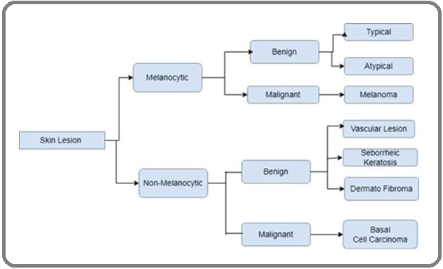
Squamous cell carcinoma, the second type, is the most common type of skin cancer and develops in thecells that make up the surface of the skin [7]. Their purpose is to provide in-depth analysis [8]. Some image processing techniques have been developed using basic research and design algorithms or systems that use methods and techniques used to solve medical problems [9]. We will search and analyze them using different parameters and extraction methods to detect skin cancer at an early stage [10]. The most dangerous type of skin cancer, melanoma, is shown in Figure 1 above. If detected early, it is treatable, but advanced melanoma is fatal [12].
Figure 1. Block Diagnosis for Skin Cancer Lesion Detection.

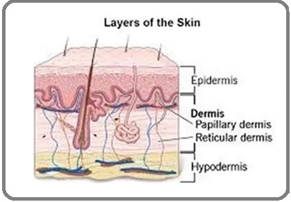
The two main layers of the skin are the epidermis and the dermis. The dermis, the word for upper layer, consists of squamous, basal and melanocyte cell layers. These cells protect the skin from invasion [14]. Neurons, blood vessels, and sweat are all found in the epidermis, sometimes called the inner layer. The three main types of cancer are melanoma, basal cell carcinoma (BCC), and squamous cell carcinoma. One of the deadliest types of cancer is melanoma [3].
Problem statement
Skin cancer is a type of tumor (cell disease) that grows in skin cells and compared to other cancers, it has affected a large number of people all over the world. An important cause of cancer is exposure to sunlight. More than 50% of the sun’s UV rays are received by the age of 24.
Skin cancer types
Below is a description of the types of skin cancer and its symptoms:
1) Basal cell carcinoma: Basal cell carcinoma usually occurs in sun-exposed areas such as the neck or face. This type of skin cancer may look like: a pus-filled or white bump on the face or a flat sore.
2) Squamous cell carcinoma: Most often, squamous cell carcinoma occurs in sun-exposed areas such as the face, ears, and hands. People with dark skin are more prone to this type of cancer in areas that are not exposed to the sun, including the legs. This type of skin cancer may look like this: a red, hard bump or a flat, scaly, crusted lesion.
3) Melanoma: Melanoma is known as one of the most malignant and deadly skin cancers worldwide. This disease is the cause of 65% of deaths related to skin cancer [7]. The importance of timely diagnosis of skin cancer is to the extent that researchers have found that skin cancer increases the probability of other cancers. Melanoma diagnosis in the first stages
Disease can significantly prevent death from this deadly cancer. In this regard, there are two major problems:
1) In most cases, due to the lack of attention of patients to skin lesions on their body surface or lack of access to experienced dermatologists, skin lesions turn from benign to malignant.
2) In many cases, skin lesions are misdiagnosed by doctors due to the similarity of their indicators. For example, melanoma and Clark are two very similar skin lesions, with the difference that melanoma is a malignant and deadly cancer and Clark is a benign skin lesion.
In the last two decades, many researches have been conducted on fast and accurate melanoma diagnosis methods based on dermatoscopic images 4 with diagnosis accuracies ranging from 70 to 95%. In general, medical images are one of the most important diagnostic tools for doctors due to the fact that they show the condition of the body in two-dimensional and even three-dimensional (by computer) [19].
Using image processing
Image processing techniques are used in many cases such as astronomy, biology, geography, archeology and in various industries such as aerospace, packaging and printing, automobile manufacturing, pharmaceuticals and medicine, electronics, food industry, steel, aluminum and copper and other industries [20, 10].
Image processing is a method of converting an image digitally and performing some operations on it, in order to get an improved image or to extract some useful information from it [11]. In a special sense, image processing is any type of signal processing where the input is an image and the output of the image processor can be an image or a set of special signs or variables related to the image. Most image processing techniques involve treating the image as a two-dimensional signal and applying standard signal processing techniques to them. Image processing often refers to digital image processing, but there are also optical and analog image processing [12.] The purpose of image processing is enhancement and improvement, recovery, pattern measurement and image recognition [21]. In the early diagnosis of cancer, image processing as a decision-making tool helps doctors. Early detection of cancer through screening images has the most important contribution in reducing the mortality caused by a specific cancer. Medical imaging plays an important role in all stages of prediction, screening, identification, staging, treatment planning, response to treatment, recurrence and palliation of cancer. In other words, images form an important part of cancer clinical protocols and have the ability to provide functional, metabolic, structural and morphological information and help in clinical decision-making along with other diagnostic tools [22].
The science of image processing has made remarkable progress in both theoretical and practical aspects in the last few decades. Image processing and analysis can be defined as a practical and technical structure to correct, increase quality and change the shape of images . Image processing basically includes capturing images with optical scanners or digital cameras and sensors, image analysis, which includes information compression, image enhancement, and pattern recognition, as well as the output stage. This stage can be an image or a report obtained from the result of image analysis [16]. Two types of methods used for image processing are analog and digital image processing. Analog techniques use image processing for hard copies such as prints and photographs, and digital image processing, which is better known today, has many applications, from the analysis of satellite images to the dimensional control of microscopic parts [17]. In the process of cancer image processing, after taking the desired image, we start with the growth of image 5 in the pre-processing stage. One of the characteristics of the images taken with a digital camera is that the brightness of the image is not uniform. This unevenness in brightness is related to a low frequency component and it is possible to correct the brightness by removing the low frequency of the image [18 , 23].
In image processing, first, pre-processing should be done on the image, which includes methods for data cleaning, consolidation, reduction and conversion. In fact, pre-processing includes the initial operations of image processing to reduce noise, change contrast and highlight the image. The image processing stage includes operations such as image segmentation, description of objects in order to facilitate image processing and classification of individual objects. In segmentation, an image is divided into areas and its constituent objects.
The next step is image segmentation, which is a necessary step in image analysis, and current techniques for image registration and description largely depend on the results of this step. The purpose of this step is to simplify or change the display of the image for more meaningful and convenient analysis. At this stage, the objects and borders of the image are defined as well as its lines and curves. In other words, this step is the process of labeling each pixel in each image, the result of which is a set of parts that together cover the entire image. Image feature extraction is another important step in the image processing process that uses algorithms to identify and isolate different features of the image, which should have sufficient information about the image. Using the results of this step, the normality or abnormality of the image is determined. By analyzing the resulting images, the doctor can identify cancer cells and provide his diagnostic results [24]. In order to diagnose melanoma, extracting appropriate features from dermatoscopic images is particularly important [25]. Feature extraction in cases such as size and color is simple. But in cases like the shape, border of lesions and tissue, it is associated with more complexity . At this stage, the clinical symptoms used by dermatologists to diagnose melanoma were selected as features, and a feature vector was assigned to each lesion. Extracting the correct features depends on the accurate demarcation of the image, which is based on the ABCD law [26]. The ABCD law examines asymmetry (A), edge (B), color (C) and diameter (D) [27]. The use of this rule makes it easy for dermatologists to diagnose melanoma, and it is very helpful in choosing the right features . After extracting the right features from the images of different skin lesions, the features were classified. To classify information, various methods can be used, including classification using neural networks, classification using the K nearest neighbor (KNN) method, and classification using the support vector machine (SVM) method [28].
Related works
Skin cancer is currently an important public health problem. The detection of skin lesions has been approached in several types of studies by various methods (Figure 2).
Figure 2. Layers of Skin.

Researchers can compare alternative methods using a variety of available data sets. Here are some studies from researchers on preprocessing and segmentation:
Their preprocessing methods include image enhancement techniques, noise removal (hairs, artifacts, and microscopic particles) from input images, and specific light correction algorithms [4]. These methods range from segmentation methods to automated skin lesion diagnosis systems.
Skin detection is based on maximum entropy threshold, feature extraction is based on grayscale appearance matrix (GLCM), and classification is based on artificial neural network (ANN). For classification, backpropagation neural network (BPN) is used [5]. This system uses a rule-based progressive sequence method to identify skin diseases.
Recommended technology users can provide relevant medical advice and diagnosis online about children’s skin conditions. Several data mining classification methods (AdaBoost, BayesNet, MLP and NaiveBayes) have been used to predict and diagnose skin conditions. Only three types of skin problems (eczema, impetigo and melanoma) improved [6]. As a clustering technique, the ABCD (Asymmetric Index Contour Color Index Diameter) approach can be used to extract features from the segment [7].
Various image processing techniques are used for image segmentation. Region expansion techniques, morphological processes, as well as adaptive thresholding and binarization were used [17]. Dermatoscopy images were segmented using the methods and results were compared with the indicated segmentation algorithm. Their accuracy is 93.71% [18].
As mentioned earlier, the use of deep convolutional networks gives more accurate results than conventional methods. an automatic detection system. First, the image undergoes a pre-processing step to reduce the shadow effect. The focus area is then precisely segmented using a segmentation algorithm that takes into account the texture and color pattern of the image. Noise and other inherent problems are then removed from the image. The watershed method is used to isolate cancerous hepatocytes. Manually detecting these problems takes a long time. The use of automated cancer detection methods enhances medical knowledge, helps diagnose diseases at an early stage and quickly yields the best results. The tools used for skin cancer detection are discussed briefly in the next section [29], along with our proposed method (Table 1).
| Ref | Techniques | Year | Results (%) |
| [8] | 19-layer deep (CNNs) | 2017 | 96.3 |
| [9] | Fully Convolution Net | 2017 | 89.7 |
| [10] | Robust Saliency-based Skin Lesion Segmentation (RSSLS) Framework | 2017 | 91.05 |
| [11] | combination of Otsu thresholding and K-means clustering | 2017 | 89.7 |
| [12] | normalizing pixels into [0–1] range, resizing, cropping | 2017 | 81.33 |
| [13] | 2D Otsu with morphological operations on color channel derived from RGB and CIELAB color space | 2016 | 93.71 |
| [14] | used a set of 84 directional filters | 2016 | 96.3 |
| [15] | Comparison Statistical region merging (SRM) Iterative stochastic, Adaptive Thresholding Color Enhancement and Iterative Segmentation Multilevel Thresholding | 2015 | 96.8 |
| [16] | 11 x 11 median filter, Gabor filter | 2015 | 94 |
| [17] | Filter adjusting the gamma values, morphological operation closing median filter | 2016 | 93.71 |
| [18] | Image enhancement, noise removal and resizing | 2015 | 96.8 |
Research Methods for Skin Cancer
Image Pre-processing
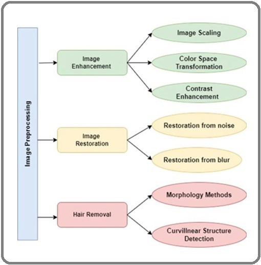
The three stages of image enhancement, image restoration and hair removal can all be used to achieve the purpose of the preprocessing steps. As shown in Figure 3, there are three types of image enhancement.
Figure 3. Image Pre-processing.

These include image enlargement, color space conversion, and contrast enhancement. Two types of image recovery are noise recovery and blur recovery. Morphological methods, curve structure detection and other methods can be used for hair removal. We need to convert the input image to the general size and color, and remove all the superfluous details like noise, bubbles, hair, etc. [24] because they come from a variety of sources.
Image segmentation
Segmentation is used to separate the ROI image from the surrounding area. We want to look at the return on investment. The result of this step helps to distinguish cancerous areas from healthy areas. Segmentation techniques can be divided into four main categories [25].
• Threshold-based - Techniques of this type include Otsu’s technique, local and global thresholding, maximum entropy, histogram-based, and others.
• Region-based – This category includes strategies such as watershed segmentation and growing seeding regions.
• Pixel-based: Techniques include fuzzy logic, random Markov fields, artificial neural networks, reinforcement learning, and more.
• Model-based: Strategies include level aggregation, deformable parametric models, and more.
Feature extraction
There are several methods used in the diagnostic procedure including.
• ABCD Rule
Asymmetrical, irregular border crossing, clinical signs of malignancy include color change (including color change within the lesion and a coloration that is different from other nevi in the patient). ), greater than 6 mm in diameter, and progressive (new or changing lesions). Later, the “sea of ugly ducklings” was developed to replace the ABCDE rule to overcome its shortcomings [27].
• Menzies method
If color homogeneity or pattern symmetry is absent, at least one of the following is present:
Pseudopods with blue and white veils, running radially and with depigmentation of wounds, broad reticulation, five to six colors, multiple blue/gray dots, and marginal black dots/globules are some examples of multiple brown dots [26].
• Seven-Point Checklist Method Unconventional pigment network, pale veil, and unusual vascular pattern (2 points each). Minor criteria (1 point each) [30] included abnormal streaks, irregular pigmentation, irregular dots/globules, and recurrent structures.
• Pattern analysis
Many knowledgeable dermatologists use pattern analysis to distinguish between benign and malignant melanocytic lesions and diagnose melanocytic lesions. Simultaneously assessing the diagnostic value of each dermoscopy feature visualized by the lesion is called pattern analysis [31].
Methods for Classifying Skin Cancer
Various methods are available to distinguish between benign and malignant melanomas. These include the use of artificial neural networks, fuzzy inference systems, and adaptive fuzzy inference neural systems as examples of artificial intelligence approaches. However, it should be noted that not all researchers opt for these classifiers. One example is the presence of a jagged band and bluish- white veil, which may indicate malignancy. Based on the angle and direction of the streaks, algorithms identify and confirm misplaced streaks. This type of detection strategy, which considers only one feature or criterion, is more accurate than machine learning techniques [32]. The upcoming discussion will focus on machine learning algorithms.
• Artificial Neural Networks
Neural networks have the ability to solve very complex problems because neurons are capable of non-linear processing. Artificial neural networks can be effectively used with medical images due to their predictive ability. Diagnosing skin cancer depends heavily on the patient’s information, but the human brain is trying to understand it. This is where ANNs play a very important role in the diagnosis of melanoma cancer and are used.
Malignant melanoma is difficult to distinguish from benign melanoma in the early stages, making skin cancer screening difficult. ANNs can solve this problem because neurons learn from examples. Using a variety of valid dermoscopic images, neurons are first trained. The back propagation method is used to train the neurons. With reverse diffusion, the current will be in the forward direction. If the output of the network is different from the desired output, the error is propagated back, which generates an error signal. In order to reduce the error, they adjust the weights. This process is repeated until the error reaches zero. The difference between the output of the network and the expected output is what is referred to as the error [33].
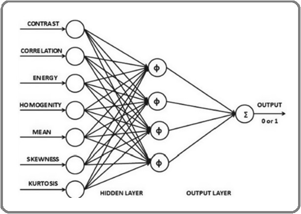
The internal dermatoscopic image functions such as curvature, mean, sprite, energy, and contrast are provided as inputs in Figure 3, and the sigmoid activation function is used to generate a zero or one output. Malignant conditions are indicated by 1 and benign conditions are indicated by zero (Figure 4).
Figure 4. Artificial Neural Network.

•Fuzzy Law Based System
After filtering, fuzzy rules are applied to the image. The result is a split image. The result image is checked to generate a binary image. The pixel values of red, green and blue components are used as fuzzy inputs. A morphological technique called closure is used to remove noise from binary images. Finally, the area filter is used to place the desired area. The next step is to evaluate the classification criteria for the retrieved ROI [34].
•Adaptive Fuzzy Inference Neural Network
AFINN combines human expert knowledge, fuzzy inference capabilities and neural network capabilities to adapt or learn the advantages of fuzzy inference rules and neural networks. As a result, our method outperforms both fuzzy logic and neural networks. To reduce the number of inputs to the AFINN system, some researchers use information acquisition strategies. AFINN has two layers: an input-output layer and a rule layer. The input and output are two components of the I/O layer. Each node in the rule layer is a fuzzy rule. The weights are fully connected from the rule layer to the output component and from the rule layer to the input part. The input part of your If-then rules contains the If part, while the rule layer stores the parts of the later part. The shape of the membership functions changes automatically during learning. AFINN changes wij and the weights are changed during the learning phase using a back-propagation method [35].
Skin cancer data sets [36]
• ISIC
• Dermofit image gallery
• PH2 data set
• MED-NODE
• Asan data sets
• Hallym dataset
• Dataset SD-198
• Dermanet New Zealand
•Derm7pt
Discussion and conclusion
In the last decade, medical imaging has been affected by the emergence of non-invasive medical tools with greater accuracy and speed. Therefore, image processing mechanisms can be widely used in many medical fields in order to improve disease identification and treatment in the early stages, without the need for invasive action.
The purpose of these mechanisms is to increase the relative quality of the information that will be extracted and interpreted by the doctor. Today, image processing is one of the growing researches that has been integrated to a great extent with the fields of medicine and biotechnology. Image processing analyzes medical images and extracts useful information from them in order to identify abnormal cases and abnormalities. Using an efficient and effective processing technique is a necessary step for the comprehensive display of clinical images that provides better diagnostic results. Since it is important to diagnose cancer in the shortest possible time, image processing mechanisms are simple and non-invasive methods for identifying cancer cells, which speed up the initial diagnosis and ultimately increase the chances of survival of cancer patients.
The purpose of this study is to check for skin cancer and effective treatments. It has the potential to revolutionize patient care, especially in improving the sensitivity and accuracy of screening for skin lesions, including early stages of skin cancer. Clinical and imaging data of all skin types are required, focusing on automatic detection of skin lesions, especially melanoma detection, by applying semantic segmentation and segmentation. from dermatoscopy images by image processing.
Acknowledgments
Statement of Transparency and Principals:
· Author declares no conflict of interest
· Study was approved by Research Ethic Committee of author affiliated Institute.
· Study’s data is available upon a reasonable request.
· All authors have contributed to implementation of this research.
References
- Automatic Skin Cancer Images Classification Elgamal M. International Journal of Advanced Computer Science and Applications (IJACSA).2013;4(3). CrossRef
- Inflammation reduces physiological tissue tolerance in the development of work-related musculoskeletal disorders Barr AE , Barbe MF . Journal of Electromyography and Kinesiology: Official Journal of the International Society of Electrophysiological Kinesiology.2004;14(1). CrossRef
- Surgical management of subungual melanoma: mayo clinic experience of 124 cases Nguyen JT , Bakri K, Nguyen EC , Johnson CH , Moran SL . Annals of Plastic Surgery.2013;71(4). CrossRef
- Anatomy, skin (integument). StatPearls [Internet] Lopez-Ojeda W, Pandey A, Alhajj M, Oakley AM . 2020.
- An Improved Segmentation Approach for Skin Lesion Classification Filali Y, Abdelouahed S, Aarab A. Statistics, Optimization & Information Computing.2019;7(2). CrossRef
- Convolutional neural networks for computer-aided detection or diagnosis in medical image analysis: An overview Gao J, Jiang Q, Zhou B, Chen D. Mathematical biosciences and engineering: MBE.2019;16(6). CrossRef
- The Development of Online Children Skin Diseases Diagnosis System Mohd Yusof M, Ab Aziz R, Fei C. International Journal of Information and Education Technology.2013. CrossRef
- Segmentation and ABCD rule extraction for skin tumors classification. arXiv preprint arXiv:2106.04372 Messadi M, Cherifi H, Bessaid A. 2021.
- Automatic Skin Lesion Segmentation Using Deep Fully Convolutional Networks With Jaccard Distance Yuan Y, Chao M, Lo Y. IEEE Transactions on Medical Imaging.2017;36(9). CrossRef
- Deep learning ensembles for melanoma recognition in dermoscopy images Codella NCF , Nguyen QB , Pankanti S, Gutman DA , Helba B, Halpern AC , Smith JR . IBM Journal of Research and Development.2017;61(4/5). CrossRef
- Saliency-Based Lesion Segmentation Via Background Detection in Dermoscopic Images Ahn E, Kim J, Bi L, Kumar A, Li C, Fulham M, Feng DD . IEEE Journal of Biomedical and Health Informatics.2017;21(6). CrossRef
- Automatic diagnosis of melanoma using linear and nonlinear features from digital image Munia TTK , Alam MN , Neubert J, Fazel-Rezai R. 2017 39th Annual International Conference of the IEEE Engineering in Medicine and Biology Society (EMBC).2017. CrossRef
- "Skin lesion classification from dermoscopic images using deep learning techniques," in Biomedical Engineering (BioMed), 2017 13th IASTED International Conference on, 2017 AR a. G.-i.-NX a. BJ a. MO Lopez . .
- "Color channel based segmentation of skin lesion from clinical images for the detection of melanoma," in Power Electronics, Intelligent Control and Energy Systems (ICPEICES), IEEE International Conference on, 2016 C. a. SLM Sagar . .
- Noninvasive Real-Time Automated Skin Lesion Analysis System for Melanoma Early Detection and Prevention O. a. BBD a. FM Abuzaghleh . IEEE Journal of Translational Engineering in Health and Medicine.2015;3. CrossRef
- “Segmentation methods for computer aided melanoma detection,” in Communication Technologies (GCCT), 2015 Global Conference, IEEE, 2015 A. a. J. R Santy . .
- “Classification of malignant melanoma and benign skin lesions: implementationof automatic ABCD rule,” R. a. M. K. Kasmi . IET Image Processing.2016;:pp. 448--455.
- Simple dynamic emission strategies for microblog filtering. In Proceedings of the 39th International ACM SIGIR conference on Research and Development in Information Retrieval (pp. 1009–1012) Tan L, Roegiest A, Clarke CL , Lin J. 2016.
- 3D ultrasound computer tomography for medical imaging Gemmeke H, Ruiter N. Nuclear Instruments and Methods in Physics Research Section A: Accelerators, Spectrometers, Detectors and Associated Equipment.2007;580. CrossRef
- Commodity cluster-based parallel processing of hyperspectral Imagery Plaza A, Valencia D, Plaza J, Martinez P. Journal of Parallel and Distributed Computing.2006;66. CrossRef
- Automatic Imaging System With Decision Support for Inspection of Pigmented Skin Lesions and Melanoma Diagnosis Alcon J, Ciuhu C, Kate W, Heinrich A, Uzunbajakava N, Krekels G, Siem D, Haan G. Selected Topics in Signal Processing, IEEE Journal of.2009;3. CrossRef
- Imaging and cancer: a review Fass L. Molecular Oncology.2008;2(2). CrossRef
- International Journal on Recent and Innovation Trends in Computing and Communication Patel S, Bhadka H, Patel B. International Journal on Recent and Innovation Trends in Computing and Communication.2014;2.
- Sine-Net: A fully convolutional deep learning architecture for retinal blood vessel segmentation Atli İ, Gedik OS . Engineering Science and Technology, an International Journal.2021;24(2). CrossRef
- The Beneficial Techniques in Preprocessing Step of Skin Cancer Detection System Comparing HoshyarAN , Al-Jumaily A, Hoshyar AN . Procedia Computer Science.2014;42. CrossRef
- The role of the ugly duckling sign in patient education Ilyas M, Costello CM , Zhang N, Sharma A. Journal of the American Academy of Dermatology.2017;77(6). CrossRef
- Mammographic resolution: influence of focal spot intensity distribution and geometry Nickoloff EL , Donnelly E, Eve L, Atherton JV , Asch T. Medical Physics.1990;17(3). CrossRef
- A Novel Method to Detect Bone Cancer Using Image Fusion and Edge Detection Nisthula P, Yadhu RB . International Journal of Engineering and Computer Science.2013;2(6):2012-2018.
- Color channel based segmentation of skin lesion from clinical images for the detection of melanoma Sagar C, Saini LM . 2016 IEEE 1st International Conference on Power Electronics, Intelligent Control and Energy Systems (ICPEICES).2016. CrossRef
- Dermoscopic evaluation of nodular melanoma Menzies SW , Moloney FJ , Byth K, Avramidis M, Argenziano G, Zalaudek I, Braun RP . JAMA dermatology.2013;149(6). CrossRef
- An estimation of the number of cells in the human body Bianconi E, Piovesan A, Facchin F, Beraudi A, Casadei R, Frabetti F, Vitale L, et al . Annals of Human Biology.2013;40(6). CrossRef
- Dermoscopy for melanoma detection in family practice Herschorn A. Canadian Family Physician Medecin De Famille Canadien.2012;58(7).
- A Non-Invasive Interpretable Diagnosis of Melanoma Skin Cancer Using Deep Learning and Ensemble Stacking of Machine Learning Models Alfi IA , Rahman MM , Shorfuzzaman M, Nazir A. Diagnostics (Basel, Switzerland).2022;12(3). CrossRef
- Applications of artificial neural networks in health care organizational decision-making: A scoping review Shahid N, Rappon T, Berta W. PloS One.2019;14(2). CrossRef
- Color Thresholding Method for Image Segmentation of Natural Images Kulkarni N. International Journal of Image, Graphics and Signal Processing.2012;4. CrossRef
- Adaptive fuzzy inference neural network Iyatomi H, Hagiwara M. Pattern Recognition.2004;37. CrossRef
License

This work is licensed under a Creative Commons Attribution-NonCommercial 4.0 International License.
Copyright
© Asian Pacific Journal of Cancer Biology , 2023
Author Details