Toward Precision Oncology in Ovarian Cancer: Dual PD-L1 and CD44 Biomarker Profiling for Recurrence Risk Assessment
Download
Abstract
Background: Epithelial ovarian carcinoma (EOC) remains one of the most fatal gynecologic cancers, often presenting at an advanced stage and prone to frequent recurrence despite aggressive treatment. Identifying reliable prognostic biomarkers is essential for improving patient stratification and optimizing adjuvant treatment strategies. This study investigated the expression patterns of programmed death-ligand 1 (PD-L1) and CD44, a recognized cancer stem cell (CSC) marker, and examined their relationships with clinicopathological features, recurrence risk, and survival outcomes in EOC.
Methods: A retrospective study was performed involving 90 patients with histologically confirmed EOC managed at National Cancer Institute, Cairo University. Immunohistochemistry was utilized to assess PD-L1 and CD44 expression in tumor samples. The associations between biomarker expression, clinical and pathological variables, recurrence-free survival (RFS), and overall survival (OS) were analyzed using Kaplan–Meier survival curves, multivariate logistic regression, and Cox proportional hazards models.
Results: PD-L1 was expressed in 42.2% of tumors and showed significant associations with advanced International Federation of Gynecology and Obstetrics (FIGO) stage (p=0.017), higher tumor grade (p=0.004), presence of distant metastasis (p=0.002), and increased recurrence rates (p<0.001). CD44 positivity was identified in 71.1% of cases, predominantly among serous carcinoma subtypes (p=0.010), and was likewise linked to a higher recurrence risk (p<0.001). Patients with PD-L1-positive tumors exhibited a notably shorter median RFS compared to PD-L1-negative patients (31.3 vs. 54.7 months, p<0.001), as did CD44-positive individuals relative to their CD44-negative counterparts (36.3 vs. 56.7 months, p<0.001). Neither biomarker, however, demonstrated a statistically significant effect on OS during a median follow-up of approximately five years. Notably, patients with concurrent PD-L1 and CD44 expression experienced the poorest RFS outcomes (mean 30.9 months), while those negative for both markers showed the most favorable prognosis (mean 56.6 months, p<0.001). Multivariate analysis confirmed PD-L1 (OR 4.86, p=0.003) and CD44 (OR 9.61, p=0.006) as independent predictors of recurrence.
Conclusions: The expression of PD-L1 and CD44 in EOC is independently associated with an elevated risk of disease recurrence, and their co-expression identifies a particularly high-risk patient subset. Although neither marker significantly influenced overall survival within the study’s follow-up period, their combined assessment holds promise for enhancing prognostic models and guiding the selection of patients for intensified monitoring and clinical trials exploring immune checkpoint blockade and CSC-targeted therapies.
Introduction
Ovarian cancer is the most fatal gynecologic malignancy and a major cause of cancer-related death in women [1, 2]. It is primarily detected in older women and has the highest fatality rate of all female reproductive malignancies. The total five-year survival rate after diagnosis is around 45%, with late-stage diagnosis dramatically decreasing the chances of survival. One of the primary causes for delayed diagnosis is the lack of early-stage symptoms, emphasizing the crucial need to understand the disease’s underlying mechanisms. Cyreductive surgery combined with platinum-based chemotherapy is the standard treatment for all stages [3, 4]. However, recurrent problems, such as cumulative toxicity, medication resistance, and recurrence after initial therapy, limit long-term success, emphasizing the critical need for novel therapeutic options.
Recent research reveals that tumor cells evade immune surveillance through a variety of methods, including the overexpression of immunological checkpoint molecules like programmed death-ligand 1 (PD-L1), which suppresses antitumor immunity. PD-L1 interacts with its receptor PD-1 on immune cells, causing T-cell fatigue and death in the tumor microenvironment (TME) and thereby enabling immunosuppression [5]. Myeloid cells are identified as essential mediators in this process. Although anti-PD-1/PD-L1 antibodies have been demonstrated to reactivate antitumor responses in various cancers, the immunological role, molecular regulation, and interactions between tumor and immune cells involving PD-L1 in ovarian cancer are poorly understood [6].
Clinical trials of PD-1/PD-L1 inhibitors in ovarian cancer have yielded modest response rates of 19% to 25% [7, 8]. Although there are transitory decreases in tumor burden, recurrence is common, and the underlying causes are not well understood. One possibility is that cancer stem cells (CSCs) have higher amounts of PD- L1. Studies in colon and breast cancers have shown that PD-L1 is preferentially expressed in CSCs rather than non-stem cancer cells, implying a relationship between CSC-associated PD-L1 and treatment resistance or relapse [9, 10].
Cancer stem cells have an important role in tumor genesis, metastasis, and heterogeneity because they are resistant to standard therapies, which may contribute to recurrence after therapy [11, 12]. Therefore, targeting CSCs is critical for attaining long-term remission. Dendritic cell vaccines, oncolytic viruses, adoptive T-cell transfer, and checkpoint inhibitors are emerging immunotherapeutic methods for the eradication of CSCs in a variety of malignancies [13]. CD44-expressing CSCs in high-grade serous ovarian cancer (HGSOC) are associated with chemoresistance and a poor prognosis [14, 15]. Similarly, higher CD44 expression in CSCs is associated with poor clinical outcomes in colorectal, breast, and ovarian malignancies [16].
Despite these developments, the mechanisms that govern CSC activation and their precise prognostic importance in ovarian cancer remain unknown [17].
The objective of this study was to evaluate the prognostic significance of programmed death-ligand 1 (PD-L1) and CD44 expression in epithelial ovarian carcinoma (EOC), with particular emphasis on their association with clinicopathological characteristics, risk of disease recurrence, and patient survival outcomes. Given accumulating evidence implicating PD-L1 in facilitating immune evasion and highlighting the role of cancer stem cells (CSCs), marked by CD44 expression, in promoting treatment resistance and disease progression, we sought to investigate the patterns of their co-expression within the tumor microenvironment. Furthermore, this study aimed to assess whether the combined expression of PD-L1 and CD44 could serve as a predictive biomarker panel for improved recurrence risk stratification and inform the development of personalized adjuvant therapeutic strategies in the management of ovarian cancer.
Materials and Methods
Patient Cohort and Sample Collection
The National Cancer Institute at Cairo University approved this retrospective investigation, which involved 90 female patients who underwent surgery and were diagnosed with ovarian cancer between 2018 and 2020. Cases without paraffin blocks or enough clinical data were excluded. Three expert pathologists examined Hematoxylin and Eosin (H&E)-stained slides to confirm tumor histological subtypes using the World Health Organization (WHO) classification. Cancer staging was calculated by combining the Tumor-Node- Metastasis (TNM) approach with the International Federation of Gynecology and Obstetrics (FIGO) criteria. Formalin-fixed, paraffin-embedded tissue blocks (FFPEs) for representative tumor tissues were chosen for immunohistochemical staining. Patient medical records and the institutional cancer registry provided relevant demographic and clinical data.
Immunohistochemistry (IHC) Procedures
For immunohistochemistry, 4-µm slices were cut from FFPE tissue blocks. Tissue sections were deparaffinized in xylene, rehydrated through graded ethanol solutions, and subjected to antigen retrieval in a high-pH solution (pH 9) using the PT LINK pre-treatment system (Agilent Technologies, Santa Clara, CA, USA; model PT100). Automated staining was performed using the VENTANA BenchMark ULTRA system (Roche Diagnostics, Basel, Switzerland; model 06522413001). The following primary antibodies were used:
• PD-L1 (clone SP142, rabbit monoclonal; Roche Diagnostics, catalog # 760-4906; ready-to-use), applied according to the manufacturer’s protocol.
• CD44 (clone SP37, rabbit monoclonal; Roche Diagnostics, catalog # 790-4436; ready-to-use).
Following peroxidase blocking and application of Protein Block, sections were incubated with primary antibodies. Each staining session included positive controls (tonsil tissue for CD44; placenta for PD-L1) and negative controls (omission of primary antibody) to ensure specificity and consistency.
Immunostaining Assessment
All slides were investigated at high magnification (400x). PD-L1 positivity was defined as membranous staining in ≥1% of tumor cells, while expression below this level was considered negative. CD44 expression was considered positive if at least 10% of tumor cells were stained in three randomly selected fields [18].
Statistical Analysis
The data were analyzed with SPSS version 27.0. Patient and tumour characteristics were summarized using descriptive statistics (means and percentages). Associations between clinicopathological factors were assessed using Pearson’s Chi-squared test or Fisher’s exact test, if appropriate. Kaplan-Meier curves were used to examine overall survival (OS) and disease-free survival (DFS), which were stratified by histological subtype, PD-L1 expression, and CD44 expression. Cox proportional hazards models were used to examine the relationship between PD-L1 expression, CD44 expression, and overall mortality, after accounting for important tumor parameters and patient age.
Results
1. Demographic and Clinicopathological Characteristics
This study included 90 patients diagnosed with epithelial ovarian carcinoma. The median age at presentation was 53 years (interquartile range: 46–60 years). Bilateral ovarian involvement was observed in 66.7% of cases, while unilateral tumors affected the right ovary in 15.6% and the left in 17.8%. Surgical management primarily involved total abdominal hysterectomy with bilateral salpingo-oophorectomy (86.7%), whereas unilateral oophorectomy was performed in 13.3% of patients.
Histologically, serous carcinoma constituted the majority (70.0%), followed by mucinous (13.3%), endometrioid (10.0%), and clear cell carcinomas (6.7%). According to the FIGO staging system, most tumors were classified as stage I (36.7%) or stage III (42.2%), with fewer cases at stage II (12.2%) or stage IV (8.9%).
High-grade tumors accounted for 64.4% of the cohort. Lymph node metastases were detected in 51.1% of patients, and lymphovascular space invasion was present in 63.3%. Only a minority (7.8%) exhibited distant metastatic spread at diagnosis, with the majority (92.2%) presenting with localized disease.
Table 1 outlines the clinicopathological features of the 90 epithelial ovarian carcinoma cases analyzed in this study, encompassing patient age, histological classification, tumor grade, FIGO staging, and recurrence status.
| Count | % | ||
| Tumor Laterality | Right ovary | 14 | 15.60 |
| Left ovary | 16 | 17.80 | |
| Bilateral ovaries | 60 | 66.70 | |
| Operation | PH | 78 | 86.70 |
| Ovariectomy | 12 | 13.30 | |
| FIGO staging | I | 33 | 36.70 |
| II | 11 | 12.20 | |
| III | 38 | 42.20 | |
| IV | 8 | 8.90 | |
| FIGO sub classification of the stage | IA | 14 | 15.60 |
| IB | 7 | 7.80 | |
| IC | 12 | 13.30 | |
| IIA | 2 | 2.20 | |
| IIB | 9 | 10.00 | |
| IIIA | 8 | 8.90 | |
| IIIB | 3 | 3.30 | |
| IIIC | 27 | 30.00 | |
| IV | 8 | 8.90 | |
| Grade | Low | 32 | 35.60 |
| High | 58 | 64.40 | |
| Histological type | Serous | 63 | 70.00 |
| Mucinous | 12 | 13.30 | |
| Clear | 6 | 6.70 | |
| Endometrioid | 9 | 10.00 | |
| PDL1 expression | Positive | 38 | 42.20 |
| Negative | 52 | 57.80 | |
| CD44 expression | Positive | 64 | 71.10 |
| Negative | 26 | 28.90 | |
| Lymphovascular emboli | Present | 57 | 63.30 |
| Absent | 33 | 36.70 | |
| Lymph node status | Positive for tumor deposits | 46 | 51.10 |
| Negative for tumor deposits | 44 | 48.90 | |
| Necrosis | Present | 54 | 60.00 |
| not present | 36 | 40.00 | |
| Metastasis | Present | 7 | 7.80 |
| Absent | 83 | 92.20 | |
| Site of Metastasis | Brain | 5 | 38.50 |
| Liver | 2 | 15.40 | |
| Pleura | 2 | 15.40 | |
| Lung | 4 | 30.80 | |
| Chemotherapy | Yes | 68 | 75.60 |
| No | 22 | 24.40 | |
| Residual disease | Macroscopic residual (R2) | 3 | 3.30 |
| Microscopic residual (R1) | 6 | 6.70 | |
| No residual (R0) | 81 | 90.00 | |
| Recurrence | Yes | 42 | 46.70 |
| No | 48 | 53.30 | |
| Status at the end of the study | Alive | 64 | 71.10 |
| Dead | 26 | 28.90 | |
| Progression | Yes | 6 | 66.70 |
| No | 3 | 33.30 | |
| Groups | CD44 or PDL1 positive | 28 | 31.10 |
| double positive | 37 | 41.10 | |
| double negative | 25 | 27.80 | |
| Group | CD44 positive | 27 | 30.00 |
| PDL1 positive | 1 | 1.10 | |
| double positive | 37 | 41.10 | |
| double negative | 25 | 27.80 |
Abbreviations, PH: Primary hysterectomy; FIGO: International Federation of Gynecology and Obstetrics; PD-L1: Programmed death-ligand 1; R0: No macroscopic residual disease; R1: Microscopic residual disease; R2: Macroscopic residual disease.
2. Immunohistochemical Expression Patterns and Correlations
PD-L1 Expression
PD-L1 immunopositivity was detected in 38 out of 90 cases (42.2%), while the remaining 52 tumors (57.8%) were negative. PD-L1 positivity showed statistically significant associations with several adverse clinicopathological features. These included advanced FIGO stage (p = 0.017), higher tumor grade (p = 0.004), increased recurrence frequency (76.3% vs. 25.0%, p< 0.001), presence of
distant metastases (18.4% vs. 0.0%, p = 0.002), and CD44 co-expression (97.4% vs. 51.9%, p< 0.001). In contrast, PD-L1 status did not significantly differ in relation to patient age, CA125 levels, tumor laterality, surgical modality, histologic subtype, lymphovascular invasion, lymph node involvement, residual disease status, administration of chemotherapy, or overall survival.
CD44 Expression
CD44 expression was observed in 64 of 90 cases (71.1%). Compared to CD44-negative tumors, CD44- positive carcinomas were significantly associated with serous histologic subtype (75.0% vs. 57.7%, p = 0.010) and increased recurrence rates (62.5% vs. 7.7%, p< 0.001). Moreover, CD44-positive patients had a markedly shorter recurrence-free survival (median RFS: 25.5 vs. 39.0 months, p = 0.037). No statistically significant differences were observed with respect to overall survival, CA125 levels, tumor grade or stage, lymph node status, or receipt of chemotherapy.
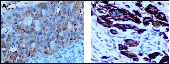
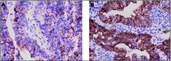
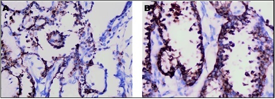
Figures 1-3 depict the immunohistochemical profiles of PD-L1 and CD44 expression within the principal histological variants of epithelial ovarian carcinoma, demonstrating notable differences in staining intensity and localization among serous, endometrioid, and clear cell subtypes.
Figure 1. Immunohistochemical Analysis of PD-L1 and CD44 in Ovarian Serous Adenocarcinoma. (A) Neoplastic cells demonstrate marked membranous and cytoplasmic positivity for PD-L1, visualized using DAB chromogen at an original magnification of ×400.(B) The corresponding section highlights diffuse and pronounced membranous expression of CD44 within tumor cells (DAB chromogen; ×400)..

Figure 2. Immunohistochemical Assessment of PD-L1 and CD44 in Ovarian Endometrioid Adenocarcinoma.(A) Tumor cells display moderate membranous staining for PD-L1, detected by DAB chromogen at ×400 magnification. (B) In the paired section, strong membranous immunoexpression of CD44 is evident in malignant cells (DAB chromogen; ×400)..

Figure 3. Immunohistochemical Characterization of PD-L1 and CD44 in Ovarian Clear Cell Adenocarcinoma.(A) Neoplastic cells reveal moderate membranous and cytoplasmic PD-L1 immunoreactivity, visualized with DAB chromogen at ×400 magnification. (B) The corresponding section demonstrates moderate to strong membranous staining of CD44 in tumor cells (DAB chromogen; ×400)..

Table 2 illustrates the immunohistochemical expression patterns of PD-L1 and CD44 across the tumor samples, categorized by histological subtype, and delineates the distribution of marker positivity within the study population.
| Variable | PDL1 Positive (n=38) | PDL1 Negative (n=52) | P value |
| Tumor Laterality | 0.093 | ||
| Right ovary | 9 (23.7) | 5 (9.6) | |
| Left ovary | 4 (10.5) | 12 (23.1) | |
| Bilateral ovaries | 25 (65.8) | 35 (67.3) | |
| Operation | 0.066 | ||
| Pan Hysterectomy | 30 (78.9) | 48 (92.3) | |
| Ovariectomy | 8 (21.1) | 4 (7.7) | |
| FIGO Stage | 0.017 | ||
| I | 8 (21.1) | 25 (48.1) | |
| II | 8 (21.1) | 3 (5.8) | |
| III | 17 (44.7) | 21 (40.4) | |
| IV | 5 (13.2) | 3 (5.8) | |
| FIGO Subclassification | 0.009 | ||
| IA | 3 (7.9) | 11 (21.2) | |
| IB | 0 (0.0) | 7 (13.5) | |
| IC | 5 (13.2) | 7 (13.5) | |
| IIA | 1 (2.6) | 1 (1.9) | |
| IIB | 7 (18.4) | 2 (3.8) | |
| IIIA | 1 (2.6) | 7 (13.5) | |
| IIIB | 2 (5.3) | 1 (1.9) | |
| IIIC | 14 (36.8) | 13 (25.0) | |
| IV | 5 (13.2) | 3 (5.8) | |
| Grade | 0.004 | ||
| Low | 7 (18.4) | 25 (48.1) | |
| High | 31 (81.6) | 27 (51.9) | |
| Histological Type | 0.173 | ||
| Serous | 28 (73.7) | 35 (67.3) | |
| Mucinous | 3 (7.9) | 9 (17.3) | |
| Clear | 1 (2.6) | 5 (9.6) | |
| Endometrioid | 6 (15.8) | 3 (5.8) | |
| CD44 Expression | 37 (97.4) | 27 (51.9) | <0.001 |
| Lymphovascular Emboli | 27 (71.1) | 30 (57.7) | 0.194 |
| Positive Lymph Node | 22 (57.9) | 24 (46.2) | 0.271 |
| Necrosis | 25 (65.8) | 29 (55.8) | 0.338 |
| Metastasis | 7 (18.4) | 0 (0.0) | 0.002 |
| Site of Metastasis† | 1 | ||
| Brain | 4 (44.4) | 1 (25.0) | |
| Liver | 1 (11.1) | 1 (25.0) | |
| Pleura | 1 (11.1) | 1 (25.0) | |
| Lung | 3 (33.3) | 1 (25.0) | |
| Chemotherapy | 31 (81.6) | 37 (71.2) | 0.256 |
| Residual Disease | 0.643 | ||
| Macroscopic residual | 2 (5.3) | 1 (1.9) | |
| Microscopic residual | 3 (7.9) | 3 (5.8) | |
| No residual | 33 (86.8) | 48 (92.3) | |
| Recurrence | 29 (76.3) | 13 (25.0) | <0.001 |
| Status at End of Study | 0.058 | ||
| Alive | 23 (60.5) | 41 (78.8) | |
| Dead | 15 (39.5) | 11 (21.2) | |
| Progression‡ | 4 (80.0) | 2 (50.0) | 0.524 |
FIGO, International Federation of Gynecology and Obstetrics; PDL1, Programmed Death-Ligand 1. Notes, † Metastatic site percentages are calculated relative to the total number of patients presenting with metastasis. ‡ Progression percentages are based on the number of patients who experienced disease progression.
3. Multivariate Predictors of Recurrence
Logistic regression analysis revealed that both PD-L1 and CD44 expressions were independent predictors of disease recurrence. PD-L1-positive tumors had an odds ratio (OR) of 4.86 for recurrence (95% confidence interval [CI]: 1.68–14.01, p = 0.003), while CD44-positive tumors conferred an even higher risk with an OR of 9.61 (95% CI: 1.92–47.98, p = 0.006).
4. Survival Analyses
Recurrence-Free Survival (RFS)
Kaplan–Meier analysis demonstrated that PD-L1 and CD44 expression were both significantly associated with shorter RFS:
• PD-L1-positive patients had a mean RFS of 31.3 months (95% CI: 24.6–38.0), significantly lower than PD-L1-negative patients at 54.7 months (95% CI: 48.0–61.4) (p< 0.001).
• CD44-positive patients exhibited a mean RFS of 36.3 months (95% CI: 30.2–42.3), compared to 56.7 months (95% CI: 52.2–61.2) in CD44-negative individuals (p< 0.001).
When stratified by combined expression, patients co-expressing both PD-L1 and CD44 had the poorest RFS (mean: 30.9 months), whereas those lacking both markers had the most favorable prognosis (mean: 56.6 months). The difference among the four combined expression groups was statistically significant (p< 0.001).
Overall Survival (OS)
In contrast to RFS, overall survival did not differ significantly between biomarker expression groups:
• PD-L1-positive vs. negative: 50.4 vs. 58.0 months (p = 0.117)
• CD44-positive vs. negative: 54.4 vs. 49.9 months (p = 0.889)
These findings suggest that while PD-L1 and CD44 expression are robust indicators of recurrence risk, they may have limited utility in predicting overall survival within the current follow-up period.
5. Progression-Free Survival (PFS)
Although not reaching statistical significance, mean PFS was numerically shorter in PD-L1-positive and CD44-positive groups:
• PD-L1-positive: 19.8 months (95% CI: 8.9–30.7)
• PD-L1-negative: 25.5 months (95% CI: 9.3–41.7),p = 0.719
• CD44-positive: 21.4 months (95% CI: 11.1–31.8)
• CD44-negative: 13.5 months (95% CI: 7.3–19.7),p = 0.968
Despite numerical trends, none of these comparisons reached statistical significance by log-rank testing.
Table 1 presents the frequencies and percentages of major clinicopathological variables and biomarker expression profiles in a cohort of 90 patients diagnosed with epithelial ovarian carcinoma. Parameters detailed include tumor laterality, type of surgical procedure performed, FIGO stage with sub-classifications, tumor grade, histological subtype, immunohistochemical expression of PD-L1 and CD44, presence of lymphovascular invasion, lymph node metastases, tumor necrosis, and distant metastatic sites. Additional data on chemotherapy administration, residual disease status following surgery, recurrence rates, survival status at the conclusion of follow-up, disease progression, and combined biomarker expression categories are also provided. All percentages reflect proportions relative to the total study population (n=90).
Table 2 provides an overview of the distribution of various clinicopathological characteristics and treatment outcomes stratified by PD-L1 expression in epithelial ovarian carcinoma cases. Parameters analyzed include tumor laterality, type of surgical procedure, FIGO stage and its subcategories, tumor grade, histopathological type, CD44 expression status, presence of lymphovascular invasion, lymph node involvement, tumor necrosis, distant metastasis (with detailed metastatic sites), chemotherapy administration, residual disease following surgery, recurrence rates, survival status at the end of follow-up, and disease progression. The p-values indicate the statistical significance of observed differences between PD-L1 positive and negative groups.
Table 3 presents a comparative overview of clinicopathological characteristics and treatment-related outcomes in patients with epithelial ovarian carcinoma, classified based on CD44 immunohistochemical expression status.
| Variable | CD44 Positive (n=64) | CD44 Negative (n=26) | P value |
| count (%) | count (%) | ||
| Tumor Laterality | 0.693 | ||
| Right ovary | 10 (15.6) | 4 (15.4) | |
| Left ovary | 10 (15.6) | 6 (23.1) | |
| Bilateral ovaries | 44 (68.8) | 16 (61.5) | |
| Operation | 0.497 | ||
| Pan Hysterectomy | 54 (84.4) | 24 (92.3) | |
| Ovariectomy | 10 (15.6) | 2 (7.7) | |
| FIGO Stage | 0.401 | ||
| I | 21 (32.8) | 12 (46.2) | |
| II | 10 (15.6) | 1 (3.8) | |
| III | 27 (42.2) | 11 (42.3) | |
| IV | 6 (9.4) | 2 (7.7) | |
| FIGO Subclassification | 0.835 | ||
| IA | 8 (12.5) | 6 (23.1) | |
| IB | 5 (7.8) | 2 (7.7) | |
| IC | 8 (12.5) | 4 (15.4) | |
| IIA | 2 (3.1) | 0 (0.0) | |
| IIB | 8 (12.5) | 1 (3.8) | |
| IIIA | 5 (7.8) | 3 (11.5) | |
| IIIB | 3 (4.7) | 0 (0.0) | |
| IIIC | 19 (29.7) | 8 (30.8) | |
| IV | 6 (9.4) | 2 (7.7) | |
| Grade | 0.714 | ||
| Low | 22 (34.4) | 10 (38.5) | |
| High | 42 (65.6) | 16 (61.5) | |
| Histological Type | 0.01 | ||
| Serous | 48 (75.0) | 15 (57.7) | |
| Mucinous | 7 (10.9) | 5 (19.2) | |
| Clear | 1 (1.6) | 5 (19.2) | |
| Endometrioid | 8 (12.5) | 1 (3.8) | |
| Lymphovascular Emboli | 42 (65.6) | 15 (57.7) | 0.479 |
| Positive Lymph Node | 33 (51.6) | 13 (50.0) | 0.893 |
| Necrosis | 38 (59.4) | 16 (61.5) | 0.849 |
| Metastasis | 7 (10.9) | 0 (0.0) | 0.103 |
| Site of Metastasis | 0.359 | ||
| Brain | 5 (45.5) | 0 (0.0) | |
| Liver | 1 (9.1) | 1 (50.0) | |
| Pleura | 2 (18.2) | 0 (0.0) | |
| Lung | 3 (27.3) | 1 (50.0) | |
| Chemotherapy | 50 (78.1) | 18 (69.2) | 0.374 |
| Residual Disease | 0.709 | ||
| Macroscopic residual | 3 (4.7) | 0 (0.0) | |
| Microscopic residual | 4 (6.3) | 2 (7.7) | |
| No residual | 57 (89.1) | 24 (92.3) | |
| Recurrence | 40 (62.5) | 2 (7.7) | <0.001 |
| Status at End of Study | 0.793 | ||
| Alive | 45 (70.3) | 19 (73.1) | |
| Dead | 19 (29.7) | 7 (26.9) | |
| Progression | 5 (71.4) | 1 (50.0) | 1 |
Abbreviations, FIGO: International Federation of Gynecology and Obstetrics; CD44: Cluster of Differentiation 44. Note: Percentages for metastatic sites are calculated relative to the number of patients within each subgroup presenting with metastases.
The analyzed parameters include tumor laterality, type of surgical intervention, FIGO staging with sub-classification, tumor grade, histological subtype, presence of lymphovascular invasion, lymph node metastasis, necrosis within the tumor, occurrence and anatomical sites of distant metastases, administration of chemotherapy, extent of residual disease following surgery, disease recurrence, survival status at the end of the study period, and documented disease progression. Data are expressed as absolute numbers and percentages within the CD44-positive and CD44-negative groups. P-values are provided to reflect the statistical significance of differences observed between the two groups.
Discussion
This investigation assessed the expression patterns of PD-L1 and CD44 in a cohort of 90 patients diagnosed with epithelial ovarian carcinoma (EOC), revealing their significant associations with unfavorable clinicopathological parameters and increased recurrence risk, although neither was predictive of overall survival (OS) within the study’s follow-up period. The median age and distribution of histological subtypes in this population were consistent with global epidemiological trends, where serous carcinoma is predominant and most commonly diagnosed in women in their fifth to sixth decades of life. Notably, over half of the cases presented at advanced FIGO stages (III–IV), and a substantial proportion demonstrated lymphovascular invasion. These findings reflect the biologically aggressive nature of serous EOC and highlight the urgent need for reliable prognostic biomarkers in this malignancy [14, 18, 19]
Approximately 42% of tumors in this cohort expressed PD-L1, with a strong correlation observed between its positivity and more advanced FIGO stage, higher tumor grade, presence of distant metastases, and increased frequency of disease recurrence. These observations support the established role of PD-L1 in mediating immune evasion in ovarian cancer, where its expression on tumor or infiltrating immune cells may suppress T-cell–mediated antitumor responses, enabling tumor progression. Although prior studies have produced mixed results regarding the relationship between PD-L1 expression and overall survival particularly in serous carcinoma subgroups where PD-L1 levels may parallel tumor-infiltrating lymphocyte (TIL) densities without necessarily predicting survival outcomes our findings reinforce its utility in identifying patients at heightened risk for early relapse rather than long-term mortality [18, 20, 21].
CD44 was expressed in 71% of the tumors examined and showed significant associations with serous histology and elevated recurrence rates. As a receptor for hyaluronan and a widely recognized marker of cancer stem cells (CSCs) in ovarian cancer, CD44 facilitates tumor initiation, chemoresistance, and metastasis. The notably shorter median recurrence-free survival (RFS) observed in CD44-positive patients in this study mirrors previous reports linking CD44 overexpression to poorer disease- free and progression-free survival outcomes in advanced EOC. These findings underscore the critical role of CSC populations in driving recurrence, even following optimal cytoreductive surgery and platinum-based chemotherapy [22-24].
Crucially, the co-expression of PD-L1 and CD44 identified a subset of patients with markedly poorer RFS compared to those lacking both markers. Multivariate logistic regression analysis confirmed both PD-L1 and CD44 positivity as independent predictors of recurrence, with particularly high odds ratios. These results suggest a synergistic effect between tumor immune evasion and CSC-associated aggressiveness in promoting relapse. Consequently, evaluating both biomarkers together could refine risk stratification models, aiding in the selection of high-risk patients for intensified surveillance protocols or enrollment in trials exploring novel combination therapies. Although both PD-L1 and CD44 demonstrated strong predictive value for RFS, neither marker significantly influenced OS within the median follow-up period of approximately five years. This lack of survival difference may reflect the impact of effective salvage therapies, such as secondary cytoreductive surgery, PARP inhibitors, and immune checkpoint inhibitors, which have improved post-recurrence outcomes in EOC. Additionally, the limited number of survival events and variability in post-relapse treatment approaches may have obscured OS differences. Nonetheless, the consistent association between these biomarkers and early relapse highlights their potential utility in guiding adjuvant treatment decisions for example, identifying candidates for trials combining immunotherapies with agents targeting CSC pathways [25, 26].
In conclusion, this study demonstrates that PD-L1 and CD44 are robust, independent indicators of recurrence risk in epithelial ovarian carcinoma, with combined expression identifying a subgroup of patients at particularly high risk for disease relapse. While neither marker predicted overall survival within the study period, their combined assessment holds promise for improving risk stratification and guiding tailored adjuvant treatment approaches, potentially incorporating combined immunotherapeutic and CSC-targeted strategies to improve long-term outcomes in this challenging malignancy.
Ethical Approval and Compliance with Standards
This research was performed in accordance with the ethical guidelines of the Declaration of Helsinki. Ethical approval was secured from the Institutional Review Board (IRB) of the National Cancer Institute, Cairo University. Patient anonymity was rigorously maintained through thorough data de-identification. Given the retrospective study design, the IRB waived the requirement for individual informed consent.
Conflict of Interest
The authors report no conflicts of interest. All elements of study design, data collection, analysis, and interpretation were independently conducted without external financial, professional, or organizational influence.
Data Availability Statement
The data underpinning this study’s results are available from the corresponding author upon reasonable request.
Funding
This research did not receive financial support from any governmental, commercial, or nonprofit funding bodies and was entirely self-funded by the authors.
References
- Global cancer statistics 2018: GLOBOCAN estimates of incidence and mortality worldwide for 36 cancers in 185 countries Bray F, Ferlay J, Soerjomataram I, Siegel RL , Torre LA , Jemal A. CA: a cancer journal for clinicians.2018;68(6). CrossRef
- Decoding β-catenin expression patterns in ovarian serous carcinoma with clinicopathological implications: insights from National Cancer Institute Ebrahim NAA , Abou-Bakr AA , Tawfik HN , Nassar HR , Adel I. Clinical & Translational Oncology: Official Publication of the Federation of Spanish Oncology Societies and of the National Cancer Institute of Mexico.2025;27(6). CrossRef
- Ovarian cancer: etiology, risk factors, and epidemiology Hunn J, Rodriguez GC . Clinical Obstetrics and Gynecology.2012;55(1). CrossRef
- The Impact of BRCA1 Expression on Survival Status in Ovarian Serous Carcinoma of Egyptian Patients Amin NH , Ahmed BA , Abou-Bakr AA , Eissa SS , Nassar HR , Gad M, Eissa M. Asian Pacific journal of cancer prevention: APJCP.2023;24(10). CrossRef
- Tumour and host cell PD-L1 is required to mediate suppression of anti-tumour immunity in mice Lau J, Cheung J, Navarro A, Lianoglou S, Haley B, Totpal K, Sanders L, et al . Nature Communications.2017;8. CrossRef
- Clinical and Prognostic Value of Antigen-Presenting Cells with PD-L1/PD-L2 Expression in Ovarian Cancer Patients Pawłowska A, Kwiatkowska A, Suszczyk D, Chudzik A, Tarkowski R, Barczyński B, Kotarski J, Wertel I. International Journal of Molecular Sciences.2021;22(21). CrossRef
- Immune checkpoint inhibitors: Key trials and an emerging role in breast cancer Gaynor N, Crown J, Collins DM . Seminars in Cancer Biology.2022;79. CrossRef
- Differential Activity of Nivolumab, Pembrolizumab and MPDL3280A according to the Tumor Expression of Programmed Death-Ligand-1 (PD-L1): Sensitivity Analysis of Trials in Melanoma, Lung and Genitourinary Cancers Carbognin L, Pilotto S, Milella M, Vaccaro V, Brunelli M, Caliò A, Cuppone F, et al . PloS One.2015;10(6). CrossRef
- Increased PD-L1 expression in breast and colon cancer stem cells Wu Y, Chen M, Wu P, Chen C, Xu ZP , Gu W. Clinical and Experimental Pharmacology & Physiology.2017;44(5). CrossRef
- Chemotherapy Induces Programmed Cell Death-Ligand 1 Overexpression via the Nuclear Factor-κB to Foster an Immunosuppressive Tumor Microenvironment in Ovarian Cancer Peng J, Hamanishi J, Matsumura N, Abiko K, Murat K, Baba T, Yamaguchi K, et al . Cancer Research.2015;75(23). CrossRef
- Cancer Stem Cells: Powerful Targets to Improve Current Anticancer Therapeutics Bighetti-Trevisan RL , Sousa LO , Castilho RM , Almeida LO . Stem Cells International.2019;2019. CrossRef
- Tumor microenvironment for cancer stem cells Kise K, Kinugasa-Katayama Y, Takakura N. Advanced Drug Delivery Reviews.2016;99(Pt B). CrossRef
- Recent Advances in Cancer Stem Cell-Targeted Immunotherapy Badrinath N, Yoo SY . Cancers.2019;11(3). CrossRef
- Prognostic impact of the cancer stem cell-related marker NANOG in ovarian serous carcinoma Lee M, Nam EJ , Kim SW , Kim S, Kim JH , Kim YT . International Journal of Gynecological Cancer: Official Journal of the International Gynecological Cancer Society.2012;22(9). CrossRef
- Cancerous ovarian stem cells: obscure targets for therapy but relevant to chemoresistance Ahmed N, Abubaker K, Findlay J, Quinn M. Journal of Cellular Biochemistry.2013;114(1). CrossRef
- Concise Review: Emerging Role of CD44 in Cancer Stem Cells: A Promising Biomarker and Therapeutic Target Yan Y, Zuo X, Wei D. Stem Cells Translational Medicine.2015;4(9). CrossRef
- CD44 expressed on cancer-associated fibroblasts is a functional molecule supporting the stemness and drug resistance of malignant cancer cells in the tumor microenvironment Kinugasa Y, Matsui T, Takakura N. Stem Cells (Dayton, Ohio).2014;32(1). CrossRef
- PD-L1 is highly expressed in ovarian cancer and associated with cancer stem cells populations expressing CD44 and other stem cell markers Alwosaibai K, Aalmri S, Mashhour M, Ghandorah S, Alshangiti A, Azam F, Selwi W, et al . BMC cancer.2023;23(1). CrossRef
- A comprehensive overview of ovarian cancer stem cells: correlation with high recurrence rate, underlying mechanisms, and therapeutic opportunities Alizadeh H, Akbarabadi P, Dadfar Al, Tareh MR , Soltani B. Molecular Cancer.2025;24(1). CrossRef
- Prognostic role of different PD-L1 expression patterns and tumor-infiltrating lymphocytes in high-grade serous ovarian cancer: a systematic review and meta-analysis Wang Y, Cai W, Xue Q, Zhang J, Zhou L, Xiong S, Deng H. Frontiers in Immunology.2023;14. CrossRef
- The emerging and challenging role of PD-L1 in patients with gynecological cancers: An updating review with clinico-pathological considerations Santoro A, Angelico G, Inzani F, Arciuolo D, Amati A, Addante F, Travaglino A, et al . Gynecologic Oncology.2024;184. CrossRef
- The Hyaluronan/CD44 Axis: A Double-Edged Sword in Cancer Cirillo N. International Journal of Molecular Sciences.2023;24(21). CrossRef
- The Role of Cancer Stem Cell Markers in Ovarian Cancer Frąszczak K, Barczyński B. Cancers.2023;16(1). CrossRef
- Role of CD44 and CD24 Expression on 2-years Disease Free Survival in Patients with Advanced Epithelial Ovarian Carcinoma Feharsal Y, Andrijono A, Singoprawiro CS , Lisnawati L, Pakasi TA , Putra AD , Kusuma F, et al . Asian Pacific journal of cancer prevention: APJCP.2024;25(2). CrossRef
- Immunotherapy in Recurrent Ovarian Cancer Chen K, Wang J, Yang N, Deng S, Sun L. Biomedicines.2025;13(1). CrossRef
- Ovarian cancer targeted therapy: current landscape and future challenges Wang G, Yang H, Wang Y, Qin J. Frontiers in Oncology.2025;15. CrossRef
License

This work is licensed under a Creative Commons Attribution-NonCommercial 4.0 International License.
Copyright
© Asian Pacific Journal of Cancer Biology , 2025
Author Details