Comparative Study of 3D Conformal Radiation Therapy by 3 Fields v/s 5 Fields Treatment Planning Techniques for Head and Neck Cancer
Download
Abstract
Objective: This study aimed to compare conventional 3-field and 5-field treatment planning techniques during 3-dimensional conformal radiotherapy (3D-CRT) in terms of organ at risk (OAR) sparing, planning target volume (PTV) coverage, treatment response, and toxicities in head and neck cancer patients.
Materials and methods: Fifty biopsy-proven and registered patients with head and neck cancer participated in this study. Patients were randomized into two arms: Arm A (3D-CRT using a 3-field delivery technique) and Arm B (3D-CRT using a 5-field delivery technique). All patients received radiation therapy on a linear accelerator with concurrent weekly cisplatin chemotherapy. Target volumes and normal structures were manually contoured on the axial slices of the planning CT scan. Patients were evaluated at the end of treatment and during follow-up visits at 1, 3, and 6 months.
Results: At the end of treatment, complete response was achieved in 22 (88%) patients in Arm A and 23 (92%) patients in Arm B. At 6 months, complete response rates were 76% and 80% in Arms A and B, respectively (p-value = 0.6836). Grade 3 xerostomia occurred in 12% of patients in Arm A and 4% of patients in Arm B (p-value = 0.92). The mean V95 (percentage of PTV receiving at least 95% of the prescribed dose) was 90.93 for the conventional 3-field technique and 93.28 for the 5-field technique (p-value = 0.08).
Conclusion: The 5-field 3D-CRT technique can effectively spare the parotid glands and other OARs while providing better PTV coverage, particularly in patients with laryngeal carcinoma, N2 or less nodal involvement, and no involvement of higher neck node levels.
Introduction
Radiation treatment of the patients with head and neck cancer is considered one of the most challenging treatments in radiotherapy because of patient anatomy, multiple targets with different dose prescriptions, extensive tumor volume and important critical organ at risk (OAR) at this site [1]. Moreover, doses up to 70-72 Gy with conventional fractionation may be prescribed. The maximal dose tolerated by the spinal cord is considered to be between 45 Gy and 50 Gy [2]. Parotid is another important OAR in head and neck radiotherapy with maximum tolerable mean dose of 26 Gy [3-4].
Intensity modulated radiation therapy (IMRT) is a refinement of three-dimensional conformal radiotherapy (3D-CRT) which allows the modulation of radiation beam intensity, So high dose can be delivered with significantly reduced dose to normal tissues [5-7]. Although IMRT is the most ideal treatment technique for head and neck cancer patients but the longer daily treatment time is a limiting factor for busy clinics. Therefore, 3DCRT is still widely used to treat HN cancers. Some modification in field can improve dose distribution so it is reasonable to consider the use of 3D-CRT for irradiation of H&N cancers as a feasible and cost-effective technique [8].
In 3D-CRT, the 3-field classic technique (two lateral opposed fields abutted to a lower anterior neck field) seems the simplest to be used. When we increase fields there can increased chances of PTV coverage and decrease dose to normal tissue.
The purpose of this article is to compare the five field 3D-CRT with classic three field 3D-CRT with the aim to determine the more effective technique for sparing parotid glands and spinal cord while keeping adequate dose coverage of PTVs.
Materials and Methods
This was a randomised prospective study conducted at Acharya Tulsi Regional Cancer Treatment and Research Institute, Sardar Patel Medical College and associated group of hospitals, Bikaner.
The study protocol includes 50 patients of histologically proven unresectable locally advanced squamous cell carcinoma of head and neck (LASCCN) of stage III-IV. Who were enrolled from April 2019 to November 2020. Inclusion criteria included inoperable, locally advanced, histologically proved stage III&IV squamous cell carcinoma of head and neck patients, ECOG performance status 0-2, Age 18-70 years, without any haematological, cardiac, renal or liver function abnormality, no previous history of treatment for the head and neck cancer and no any other concurrent malignancies. Total 50 patients, randomly selected were divided into two groups of 25 patients in each; randomization was done using computer software. (https://www.randomizer.org/). Both arms were irradiated by linear accelerator (Make:Varian, Model: 2300CD with multileaf collimators having 40 pairs of leaves and each leaf having 1cm width at isocentre) with concurrent chemotherapy in form of three weekly Cisplatin. CT imaging was done for each patient prior to start of the treatment. All patients were undergo head-and-neck immobilization with a thermoplastic mask and CT simulation according to standard protocols. Target volumes and normal structures were be manually contoured on the axial slices of the planning CT scan. In this study, the total dose of up to 70Gy was prescribed to the PTV, with the conventional fractionation scheme arms were compared with the dose prescription of 44 Gy. Patients were evaluated at end of treatment, 1st, 3rd & 6thmonth follow up visits by complete clinical examination including laryngoscopy (Direct & Indirect), haematological investigation, USG abdomen, chest X- ray & soft tissue neck X-ray will also be done on follow up visits. CECT head and neck was done at 3rdand 6th months follow-up visits.
Results
Table 1 shows patients characteristics, which are comparable in both arms.
| Patients characteristics | Study Arm | Control Arm |
| Age (in years) | ||
| Median age | 56yr | 56 yr |
| Range | 38-70 yrs | 36-69 yrs |
| Sex | ||
| Male | 24 | 23 |
| Female | 1 | 2 |
| ECOG | ||
| 0 | 9 | 9 |
| 1 | 14 | 13 |
| 2 | 2 | 3 |
| Tumor stage | ||
| T2 | 1 | 2 |
| T3 | 19 | 18 |
| T4 | 5 | 5 |
| Nodal stage | ||
| N0 | 6 | 12 |
| N1 | 7 | 6 |
| N2 | 12 | 5 |
| N3 | 0 | 2 |
| Group stage | ||
| Stage III | 11 | 15 |
| Stage IV | 14 | 10 |
| Anatomical site | ||
| Oral cavity/ Oropharynx | 13 | 17 |
| Hypopharynx | 8 | 5 |
| Larynx | 4 | 3 |
Table 2 shows response evaluation for disease control.
| 3 field 3D-CRT arm | 5 field 3D-CRT arm | |||||||
| Disease Response | End of treatment | At 1 month after treatment | At 3 months after treatment | At 6 month after treatment | End of treatment | At 1 month after treatment | At 3 month after treatment | At 6 months after treatment |
| CR | 22 | 21 | 20 | 19 | 23 | 22 | 21 | 20 |
| PR | 1 | 2 | 2 | 2 | 1 | 2 | 1 | 2 |
| SD | 1 | 1 | 0 | 0 | 0 | 0 | 0 | 0 |
| PD | 1 | 1 | 2 | 2 | 1 | 1 | 2 | 2 |
| DEATH | 0 | 0 | 1 | 2 | 0 | 0 | 1 | 1 |
| Total | 25 | 25 | 25 | 25 | 25 | 25 | 25 | 25 |
The response evaluation was done according to the RECIST criteria (Response Evaluation Criteria in Solid Tumors v-1.1) at the end of the treatment, one, three and six months after end of treatment. At the end of treatment, 22 patients showed CR, 1 patient showed PR, 1 patient showed SD and 1 patient was in progression of disease in control arm; While in study arm 23 patients had CR, 1 patient had PR and one patient had PD.
After 6 months of completion of treatment, Total 19 vs 20 patients had CR, 2 patients in each arm had PR and 2 patients in each arm had PD in control and study arms respectively (p value=0.6836).
After 6 months total 3 patients were expired in control arm and two patients expired in study arm due to disease itself. The patients who have partial response after 3 months treated with chemotherapy.
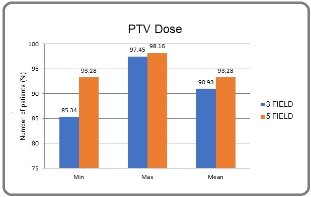
Figure 1 shows mean PTV dose coverage, V95 and max PTV dose were compared in between convention 3 field technique vs 5 field technique.
Figure 1. PTV Coverage.

Chi square test and p value are used for statistical analysis. Mean V95 was 90.93 with conventional 3 field technique and was 93.28 with 5 field technique. There is no significant difference between the mean V95 with the conventional technique or with 5 field technique (90.93% vs. 93.28%; p = 0.08). Absolute Mean PTV dose coverage do not differ significantly among the 3 field 3D-CRT arm and 5 field 3D-CRT arm (89% vs 93.36 % [p=0.073]). Mean of maximum dose coverage was also similar between two techniques. Mean of maximum dose was 109.62% vs 109.71% (p = 0.076).
Table 3 shows that, maximum doses did not differ significantly among the two techniques. Mean maximum spinal cord in conventional 3 filed vs 5 field is 45.15 Gy vs 44.83 Gy. None of the patient in either arm had received more than 48 Gy dose to spinal cord. These doses are well within the dose constrain of spinal cord.
| DOSE (Gy) | 3 field 3D-CRT arm | 5 field 3D-CRT arm |
| Less than 44Gy | 4 | 8 |
| 44.01 – 44.99 | 3 | 5 |
| 45- 45.99 | 11 | 6 |
| 46-46.99 | 5 | 4 |
| 47-47.99 | 2 | 2 |
| More than 48 Gy | 0 | 0 |
| Total | 25 | 25 |
Table 4 suggests than Mean parotid dose (MPD) in conventional 3 field technique is 34.11 Gy and 34.80 Gy in right and left parotid respectively.
| 3 field 3D-CRT | 5 field 3D-CRT | |
| Right MPD (cGy) | 3411.43 | 3129.16 |
| Left MPD (cGy) | 3480.72 | 3203.36 |
Similarly MPD with 5 field technique is 31.29 Gy and 32.03 Gy in right and left sided parotid respectively. MPD with 5 field technique is reduced in comparison to conventional 3 field technique, although it was statistically insignificant (p value 0.92). In this study, maximum doses did not differ significantly among the two techniques. Mean maximum spinal cord in conventional 3 filed vs 5 field is 45.15 Gy vs 44.83 Gy. None of the patient in either arm had received more than 48 Gy dose to spinal cord. These doses are well within the dose constrain of spinal cord (Table 4).
Mean parotid dose (MPD) in conventional 3 field technique is 34.11 Gy and 34.80 Gy in right and left parotid respectively. Similarly MPD with 5 field technique is 31.29 Gy and 32.03 Gy in right and left sided parotid respectively. MPD with 5 field technique is reduced in comparison to conventional 3 field technique, although it was statistically insignificant (p value 0.92).
Discussion
Head and neck cancer treatment is the most difficult to plan because of patient anatomy, multiple targets with different dose prescriptions, large extension of the treatment region, and the number of structures at risk. To overcome planning difficulties, highly sophisticated techniques such as IMRT, IMAT, or VMAT have been developed, which yield much better results than does three- dimensional conformal radiotherapy (3DCRT), especially in the sparing of the organs-at-risk (OARs) (Table 5).
| S.N. | Study | Purpose | Rseult |
| 1 | Antonella Fogliata, et al 1999 [9] | Compare three field vs five field treatment technique | PTV coverage 95.6 in 5 field,98.7 in 3 field, less toxicity in 5 field |
| 2 | Lee N. et al 2004 [10] | FPMS technique | 59.4 Gy at 75% isodose curve |
| 3 | Portaluri.et al 2006 [11] | FIF in 3D-CRTvs IMRT | PTV coverage was 96.2 in FIF, mean dose to parotid 38.3Gy |
| 4 | Mohmed Yassine Herrasi at al 2013 [12] | FPMS/3-DCONPAS/FIF/BELLINZONA | PTV coverage better in FPMS and FIF technique |
However, these techniques cannot be universally used, due to unavailability of adequate equipment, organization, or patient status. In this study, we tried to study that can 5 field instead of 3 field technique can improve PTV coverage while sparing organ at risk such as parotid, spinal cord. In this study PTV coverage is improved with 5 field technique although it was statistically insignificant. Mean parotid dose is also reduced with 5 filed technique but it was higher than recommended dose constraints of MPD (mean parotid dose). However, in analysis of larynx subgroup especially with less than N2 and not involving higher node, MPD was nearer to target dose constrain of 26 Gy. None of patient in either arm had received radiation dose higher than recommended dose constrain of spinal cord, brain stem and lens.
In conclusion, 5 field 3 D-CRT technique can be used to spare parotid and other OAR and better PTV coverage specially in larynx carcinoma patients with N2 or less nodal involvement and not involving higher neck node levels.
Limitations
Small size of sample and short follow-up of patients is limitation of this study. Study with larger sample size and of longer follow-up will help to confirm the results of this study.
References
- Spinal cord dose from standard head and neck irradiation: implications for three-dimensional treatment planning Martel MK , Eisbruch A, Lawrence TS , Fraass BA , Ten Haken RK , Lichter AS . Radiotherapy and Oncology: Journal of the European Society for Therapeutic Radiology and Oncology.1998;47(2). CrossRef
- Treatment planning constraints to avoid xerostomia in head-and-neck radiotherapy: an independent test of QUANTEC criteria using a prospectively collected dataset Moiseenko V, Wu J, Hovan A, Saleh Z, Apte A, Deasy JO , Harrow S, et al . International Journal of Radiation Oncology, Biology, Physics.2012;82(3). CrossRef
- Xerostomia after radiotherapy. What matters--mean total dose or dose to each parotid gland? Tribius S, Sommer J, Prosch C, Bajrovic A, Muenscher A, Blessmann M, Kruell A, et al . Strahlentherapie Und Onkologie: Organ Der Deutschen Rontgengesellschaft ... [et Al].2013;189(3). CrossRef
- International Commission on Radiation Units and Measurements Menzel HG . Journal of the ICRU.2014;14(1). CrossRef
- Dose, volume, and function relationships in parotid salivary glands following conformal and intensity-modulated irradiation of head and neck cancer Eisbruch A, Ten Haken RK , Kim HM , Marsh LH , Ship JA . International Journal of Radiation Oncology, Biology, Physics.1999;45(3). CrossRef
- A prospective study of salivary function sparing in patients with head-and-neck cancers receiving intensity-modulated or three-dimensional radiation therapy: initial results Chao KS , Deasy JO , Markman J, Haynie J, Perez CA , Purdy JA , Low DA . International Journal of Radiation Oncology, Biology, Physics.2001;49(4). CrossRef
- The potential for sparing of parotids and escalation of biologically effective dose with intensity-modulated radiation treatments of head and neck cancers: a treatment design study Wu Q, Manning M, Schmidt-Ullrich R, Mohan R. International Journal of Radiation Oncology, Biology, Physics.2000;46(1). CrossRef
- ConPas: a 3-D conformal parotid gland-sparing irradiation technique for bilateral neck treatment as an alternative to IMRT Wiggenraad R, Mast M, Santvoort J, Hoogendoorn M, Struikmans H. Strahlentherapie Und Onkologie: Organ Der Deutschen Rontgengesellschaft ... [et Al].2005;181(10). CrossRef
- Critical appraisal of a conformal head and neck cancer irradiation avoiding electron beams and field matching Fogliata A, Cozzi L, Bieri S, Bernier J. International Journal of Radiation Oncology, Biology, Physics.1999;45(5). CrossRef
- A forward-planned treatment technique using multisegments in the treatment of head-and-neck cancer Lee N, Akazawa C, Akazawa P, Quivey JM , Tang C, Verhey LJ , Xia P. International Journal of Radiation Oncology, Biology, Physics.2004;59(2). CrossRef
- Three-dimensional conformal radiotherapy for locally advanced (Stage II and worse) head-and-neck cancer: dosimetric and clinical evaluation Portaluri M, Fucilli FIM , Castagna R, Bambace S, Pili G, Tramacere F, Russo D, Francavilla MC . International Journal of Radiation Oncology, Biology, Physics.2006;66(4). CrossRef
- Comparative study of four advanced 3d-conformal radiation therapy treatment planning techniques for head and neck cancer Herrassi MY , Bentayeb F, Malisan MR . Journal of Medical Physics.2013;38(2). CrossRef
License

This work is licensed under a Creative Commons Attribution-NonCommercial 4.0 International License.
Copyright
© Asian Pacific Journal of Cancer Care , 2024
Author Details
How to Cite
- Abstract viewed - 0 times
- PDF (FULL TEXT) downloaded - 0 times
- XML downloaded - 0 times