Clinicopathological Profile of Sinonasal Masses: A Hospital Based Study
Download
Abstract
Objective: To analyze the clinicopathological spectrum of sinonasal masses, encompassing both neoplastic and non-neoplastic lesions, based on clinical, radiological, and histopathological findings.
Methods: A retrospective study was conducted over a period of three years at Fakhruddin Ali Ahmed Medical College, Assam. A total of 50 patients presenting with sinonasal masses were evaluated. The assessment included clinical examination, radiological investigations, and histopathological evaluation, with immunohistochemistry applied in select cases for diagnostic confirmation.
Results: Non-neoplastic lesions were the most common, with inflammatory polyps accounting for the majority. Among the neoplastic lesions, inverted papilloma was the most frequent benign tumour, while squamous cell carcinoma was the predominant malignant neoplasm.
Conclusion: Sinonasal masses exhibit a wide pathological spectrum. Histopathological examination remains the gold standard for definitive diagnosis, with immunohistochemistry serving as a useful adjunct in certain cases.
Introduction
Sinonasal masses encompass a broad spectrum of lesions originating in the nasal cavity and paranasal sinuses, comprising non-neoplastic and neoplastic (benign and malignant) masses.
The nasal cavity contains different types of epithelial (squamous, neural, olfactory) and mesenchymal (bone, cartilage, muscle and vascular) tissues. Lesions may arise from any of these types of tissues [1]. They are exposed to a variety of infections and environmental influences giving rise to various infections, tumor-like lesions and true neoplasms. As a result of these multifaceted exposures, various inflammatory conditions, infections and neoplasms can occur in the sinonasal tract [2].
Sinonasal malignancies make up <5% of all head and neck neoplasms [3].
The overlap in clinical presentations nasal obstruction, rhinorrhea, epistaxis, and facial swelling often complicates differential diagnosis. While imaging modalities like CT and MRI provide structural details, histopathological examination (HPE) remains the gold standard for identifying non-neoplastic, benign and malignant neoplastic lesions.
This study aims to comprehensively analyze sinonasal masses in a tertiary care hospital, highlighting diagnostic challenges and emphasizing the role of histopathology inaccurate classification.
Aims and Objectives
1. To analyze the clinical and histopathological spectrum of sinonasal masses.
2. To classify lesions as non-neoplastic or neoplastic (benign or malignant) based on histopathological features.
3. To identify diagnostic challenges in evaluating sinonasal masses.
Materials and Methods
A retrospective study of 50 cases of sinonasal masses was conducted from August 2021 to July 2024 at Fakhruddin Ali Ahmed Medical College, Assam.
All patients with clinically diagnosed sinonasal masses during the study period were included.
Clinical history and imaging results (CT/MRI) were recorded from the data archives.
Biopsy specimens sent to the Department of Pathology were processed, sectioned, and stained with hematoxylin and eosin (H&E).
Immunohistochemistry (IHC) was performed for selected malignant cases.
Collected data were studied, analyzed and presented in tabulated forms.
Results
Age and Gender Distribution
As dwpicted in Tabe 1, the age range of patients was 9–90 years, with a peak incidence in the 16–30 years age group. A male predominance was observed with a male- to-female ratio of 1.6:1.
| Age Group (in years) | Male | Female | Total (Percentage) |
| 0-15 | 8 | 3 | 11 (22) |
| 16-30 | 8 | 5 | 13 (26) |
| 31-45 | 7 | 3 | 10 (20) |
| 46-60 | 6 | 6 | 12 (24) |
| 61-75 | 2 | 1 | 03 (06) |
| 76-90 | 0 | 1 | 01 (02) |
Clinical Presentation
The various clinical presentations are shown in Table 2.
| Mode of Presentation | No. of Cases | Percentage |
| Nasal obstruction | 30 | 60 |
| Nasal discharge (Rhinorrhea) | 18 | 36 |
| Nasal mass | 14 | 28 |
| Epistaxis | 8 | 16 |
| Headache | 6 | 12 |
| Facial swelling | 4 | 8 |
The most common presenting symptom was nasal obstruction (60%), followed by rhinorrhea (36%), epistaxis (20%), and facial swelling (10%).
Histopathological Distribution
The histopathological distributions are depicted in Table 3, 4 and 5 respectively.
| Type of Lesion | NO. oF Cases | Percentage |
| Non-Neoplastic | 36 | 72 |
| Neoplastic-Benign | 9 | 1 |
| Neoplastic-Malignant | 5 | 10 |
| Diagnosis | NO. of Cases | Percentage |
| Inflammatory polyp | 29 | 58 |
| Fungal sinusitis | 3 | 6 |
| Pyogenic granuloma | 2 | 4 |
| Rhinosporidiosis | 1 | 2 |
| Fibrous dysplasia | 1 | 2 |
| Diagnosis | No. of Cases | Percentage |
| Benign | ||
| Inverted papilloma | 5 | 10 |
| Haemangioma | 2 | 4 |
| Juvenile angiofibroma | 1 | 2 |
| Ameloblastoma | 1 | 2 |
| Malignant | ||
| Squamous cell carcinoma | 3 | 6 |
| Ewings sarcoma | 1 | 2 |
| Melanoma | 1 | 2 |
The majority of cases (72%) were non-neoplastic, predominantly inflammatory polyps. Neoplastic lesions accounted for 26% of cases, with 18% benign and 10% malignant.
Discussion
In the present study, 50 cases of sinonasal mass were included retrospectively.
The age distribution ranged from 9yrs to 90 yrs with most of the cases being in the age group 16-30 yrs. There was a male preponderance with 31 males and 19 females. Male to female ratio being 1.6:1, similar to a study conducted by N Khan et al [4].
Most common presenting symptom was nasal obstruction and stuffiness (60% of the cases) followed by nasal discharge (36% of the cases). This is similar to study conducted by P. Agarwal et al in 2016 [5]. Other and nasal mass.
Majority of the patients presented within 1 year of duration of onset of symptoms, earliest being 4 months. Some of the patients had undergone varied treatment in the periphery as the symptoms were not severe, leading to their late presentation to our hospital. Out of the 50 cases in our study, 36 (72%) cases were histopathologically diagnosed as non-neoplastic, and 14 cases were put in the neoplastic category.
The neoplastic were further classified into benign and malignant, with 9 (18%) and 5 (10%) cases respectively. This findings were similar to studies conducted by A Lathi et al [6] and P. Agarwal et al [5].
In the non-neoplastic group, most common histopathological diagnosis was of inflammatory polyp that is 29 cases out of 50 (58%). This is in concordance with studies conducted by S. S. Bist et al [7], and N. Khan et al [4].
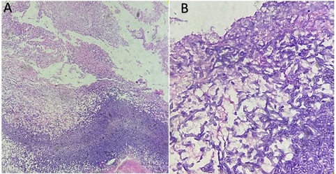
Second most common non-neoplastic diagnosis was fungal sinusitis (6%). One case of invasive aspergillosis was found (Figure 1).
Figure 1. H and E Picture Showing Aspergillosis (A) 10X, (B) 40X.

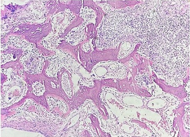
Other non-neoplastic lesions included cases of rhinosporidiosis, pyogenic granuloma and Fibrous dysplasia (Figure 2).
Figure 2. H and E Stained Picture Showing Fibrous Dysplasis (10X).

Of the neoplastic benign cases, inverted papilloma was the most common diagnosis (5 cases; 10% of the total). This is similar to study conducted by Bakari et al [8] and Kumari et al [9].
But these findings were discordant with those found by Bist et al [7], Patel et al [1] and N. Khan et al [4], where the most common benign neoplasms were angiofibroma and haemangioma, respectively.
One single case of juvenile angiofibroma was found in a 14 yr old male. Variable wall thickness with staghorn blood vessels and stellate cells mixed in loose stroma was seen on Histopathological examination.
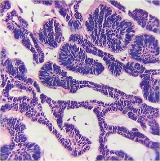
One single case of ameloblastoma was found in a 45 yr old male. Tissue biopsy was received first from the maxillary sinus, followed by entire resected mass (partial maxillectomy) for histopathology which confirmed the case as ameloblastoma (Figure 3).
Figure 3. HPE Showing Picture of Ameloblastoma.

Tumors that grow in the maxilla may secondarily extend through the nasal and paranasal cavities or it can be primary. Primary ameloblastomas of sinonasal tract, without connection with gnathic areas, are rare [10].
Of the 5 malignant neoplasms found in our study, we had three cases of Squamous cell carcinoma, one case of Ewings sarcoma and Mucosal Melanoma each.
These presented with nasal mass, discharge, epistaxis and facial swelling. All of the cases were male with age ranging from 28 to 60 years. Maxillary sinus was most commonly involved site. This is similar to the findings by Satarkar et al [11] and A Lathi et al [6].
In studies conducted by Patel et al [1] and P. Agarwal et al [6] the most common malignancy found in the sinonasal area was squamous cell carcinoma which is similar to our study. Diagnosis of squamous cell carcinoma was done on histopathological examination.
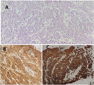
A 32 yr old male clinically presented with a right maxillary sinus mass. CT showed an enhancing soft tissue mass without calcification. On histopathological examination it was diagnosed as Round cell tumor of the maxilla. On immunohistochemistry positivity for CD99, NKX2.2 and synaptophysin confirmed it as Ewing’s sarcoma (Figure 4).
Figure 4. Ewing Sarcoma (A): HPE on H and E stain, (B) Positive CD99 membranous expression, (C) Positive NKX2.2 Nuclear expression.

Ewing Sarcoma of the sinonasal tract is a rare entity with high mortality, but few standardized treatment protocols exist [12]. Multidisciplinary approach is of utmost importance for early diagnosis and treatment.
Another case of a 60yr old female with right sided nasal cavity mass, revealed small round cells around vessels, with hemorrhage and pigment-laden cells on biopsy from the mass. MRI showed a large irregular infiltrative heterogeneously enhancing multilobulated lesion with epicenter involving right nasal cavity and right maxillary sinus. On immunohistochemistry weak S-100 positivity and strong HMB45 positivity was seen, confirming it as malignant melanoma (Figure 5).
Figure 5. Malignant Melanoma: (A) HPE on H and E stain; (B) HMB45 cytoplasmic expression; (C) S100 cytoplasmic and nuclear expression.

Sinonasal mucosal melanoma accounts for 1% of all the melanomas encountered and 4% of all the sinonasal tumors [13]. It is highly aggressive due to its rich vascularity and tends to metastasize. It poses a diagnostic challenge as around 20% are multifocal and around 40% are amelanotic [14]. In conclusion, the initial symptoms presented by patients with sinonasal masses are often indistinguishable from one another, making it challenging for appropriate diagnosis.To ensure a precise diagnosis, healthcare professionals should employ a multifaceted approach that includes not only clinical assessments but also detailed radiological imaging and histopathological analysis aided by immunohistochemical studies wherever necessary.These combined diagnostic tools are essential for accurately identifying the type of sinonasal mass and determining the most appropriate treatment strategy.
Acknowledgements
Funding Statement
There is no specific grant from funding agencies in the public, commercial, or not-for-profit sectors.
Scientific approval
The study was approved by Institutional Ethics Committee; FAAMCH Medical Ethics Committee.
Ethical Declaration
The authors declare that they have no known competing financial interests or personal relationships that could have appeared to influence the work reported in this paper
References
- Clinicopathological study and management of masses in the sinonasal cavity and nasopharynx: a case series of 42 cases Patel U, Chauhan H, Patel N. The Egyptian Journal of Otolaryngology.2023;39(1). CrossRef
- Histopathological study of lesions of nasal cavity and paranasal sinuses Kulkarni A, Shetty A, Pathak P. Indian Journal of Pathology and Oncology.2020;7(1):88-93.
- The contemporary management of cancers of the sinonasal tract in adults Thawani R, Kim MS , Arastu A, Feng Z, West MT , Taflin NF , Thein KZ , et al . CA: a cancer journal for clinicians.2023;73(1). CrossRef
- Masses of nasal cavity, paranasal sinuses and nasopharynx: A clinicopathological study Khan N., Zafar U., Afroz N., Ahmad S. S., Hasan S. A.. Indian Journal of Otolaryngology and Head and Neck Surgery: Official Publication of the Association of Otolaryngologists of India.2006;58(3). CrossRef
- Sinonasal Mass-a Recent Study of Its Clinicopathological Profile Agarwal P., Panigrahi R.. Indian Journal of Surgical Oncology.2017;8(2). CrossRef
- Clinico-pathological profile of sinonasal masses: a study from a tertiary care hospital of India Lathi A., Syed M. M. A., Kalakoti P., Qutub D., Kishve S. P.. Acta Otorhinolaryngologica Italica: Organo Ufficiale Della Societa Italiana Di Otorinolaringologia E Chirurgia Cervico-Facciale.2011;31(6).
- Clinico-pathological profile of sinonasal masses: An experience in tertiary care hospital of Uttarakhand Bist S. S., Varshney S, Baunthiyal V, Bhagat S, Kusum A. National Journal of Maxillofacial Surgery.2012;3(2). CrossRef
- Clinico-pathological profile of sinonasal masses: an experience in national ear care center Kaduna, Nigeria Bakari A, Afolabi OA , Adoga AA , Kodiya AM , Ahmad BM . BMC research notes.2010;3. CrossRef
- Clinicopathological Challenges in Tumors of the Nasal Cavity and Paranasal Sinuses: Our Experience Kumari S, Pandey S, Verma M, Rana AK , Kumari S. Cureus.2022;14(9). CrossRef
- Ameloblastoma of the sinonasal tract: report of a case with clinicopathologic considerations Tranchina MG , Amico P, Galia A, Emmanuele C, Saita V, Fraggetta Fi. Case Reports in Pathology.2012;2012. CrossRef
- Tumors and tumor-like conditions of the nasal cavity, paranasal sinuses, and nasopharynx: A study of 206 cases Satarkar R. N., Srikanth S.. Indian Journal of Cancer.2016;53(4). CrossRef
- Sinonasal Ewing Sarcoma: A Case Report and Literature Review Lin JK , Liang J. The Permanente Journal.2018;22. CrossRef
- Mucosal melanoma of the head and neck Ascierto PA , Accorona R, Botti G, Farina D, Fossati P, Gatta G, Gogas H, et al . Critical Reviews in Oncology/Hematology.2017;112. CrossRef
- Nasal mucosal melanoma Wahid NW , Meghji S, Barnes M. The Lancet. Oncology.2019;20(5). CrossRef
License

This work is licensed under a Creative Commons Attribution-NonCommercial 4.0 International License.
Copyright
© Asian Pacific Journal of Cancer Care , 2025
Author Details
How to Cite
- Abstract viewed - 0 times
- PDF (FULL TEXT) downloaded - 0 times
- XML downloaded - 0 times