Refractory Craniopharyngioma: Successful Treatment with LINAC Based Stereotactic Radiosurgery (SRS): First Case in Nepal
Download
Abstract
Purpose: To present first case of refractory craniopharyngioma treated successfully with SRS in Nepal.
Bckground: Craniopharyngioma is a benign tumour, which progresses slowly and compresses the pituitary gland and nearby structures. First line of treatment is surgery followed by adjuvant radiotherapy as complete resection is usually not feasible. Here, we are reporting a case of recurrent craniopharyngioma treated with LINAC based SRS.
Case Presentation: A 43 years old man diagnosed case of craniopharyngioma in May 2019. He underwent left pterional craniotomy and subtotal resection of tumour and kept on observation. He developed symptomatic as well as radiological recurrence in July 2020. Second debulking was not possible, so we did SRS on 23rd August 2020; 14Gy Dose was delivered to gross tumour volume. Six months after SRS, Patient is doing well.
Conclusions: LINAC based SRS is a frameless, non-invasive and safe procedure with excellent clinical outcomes for recurrent or residual craniopharyngioma.
Introduction
Craniopharyngiomas are benign tumours arising along the craniopharyngeal ducts. There are two histological subtypes – Adamatnomatous and Papillary Craniopharyngioma. Adamatnomatous have bimodal peak of incidence i.e. 5-15 years and 45-60 years whereas Papillary are mainly found in 5th and 6th decades of Life [1]. Usual presenting symptoms include lethargy, confusion, anorexia, nausea, vomiting and bitemporal hemianopia, papilledema and horizontal double vision [2-4]. Endocrinological, ophthalmological and neuropsychological assessment should be done at diagnosis [4].
The standard of care for craniopharyngioma is resection with or without adjuvant external Beam Radiotherapy (EBRT). Complete resection is the goal but it is not feasible most of the times due to associated morbidity [5]. Sub Total Resection (STR) is associated with high rate of recurrence, so, adjuvant radiotherapy is an integral part of treatment which reduces progression by 75 – 90 % [6]. Recurrent tumours are treated with second debulking surgery and EBRT.
Case Report
A 43 years old male, presented with symptoms like irritability, mood swings and headache in May 2019. Symptoms were of insidious onset and gradually progressive in nature. He was seen by general physician and was later referred for neurosurgical consultation. MRI brain was suggestive of mass in the sellar region.
He underwent left pterional craniotomy and subtotal resection of tumour. He was kept on close follow up. He was free of symptoms for 1 year when he suddenly developed headache, confusion, nausea and loss of memory, for which he presented to our hospital. We did further evaluation with MRI Brain and it was suggestive of post-operative changes in sella and suprasellar region, with 20 mm × 14 mm lesion in suprasellar cistern. The lesion was extending to floor of third ventricle, suggestive of recurrent lesion. He was planned for re-excision and neurosurgery consultation was sought. After thorough evaluation, Neurosurgeon termed it as inoperable and was referred back to us for External Beam Radiotherapy. So we discussed different RT techniques like conventional fractionation schedule, stereotactic Radiotherapy and Stereotactic surgery with patient’s attendants. They opted for stereotactic radiosurgery (SRS) treatment. After taking informed written consent, he was treated with SRS on 23 August 2020 with a dose of 14Gy to Gross Tumour Volume. After 6 months post SRS, he is completely free of symptoms and MRI in February 2021 is suggestive of significant reduction in size of the lesion.
Procedural techniques
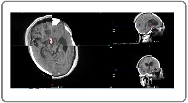
After obtaining informed written consent, patient was taken for CT simulation. It was done with Siemens Somatom Definition AS+ 4D CT simulator. Patient was made to lie down on CT Couch in head first supine position with slight chin up. For strict immobilization, frameless stereotactic Double Shell Positioning System (DSPS) was used. CT scans with slice thickness of 1 mm were acquired. Images were imported to Eclipse Treatment Planning System (TPS) of Varian True Beam System version 15.6. Recent MRI images were registered and matching was done with planning CT images (T1 Contrast with Contrast CT) (Figure 1).
Figure 1. Showing the Fusion of MRI Images with CT for Delineation of Target.

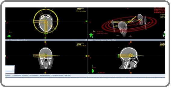
Radiation oncologist and neuro-radiologist delineated the gross tumour volume (GTV) on CT images and was verified with registered MRI images. Organs at risk contoured were Brainstem, Optic Chiasm and Optic nerves. The dose prescribed to Planning Target Volume (1 mm margin to GTV) was 14Gy in single exposure. For Planning, rapid arc inverse treatment technique was used with 6 coplanar arcs (3 partial arcs and 3 full arcs) (Figure 2).
Figure 2. Showing 6 Coplanar Arcs Used for this Patient.

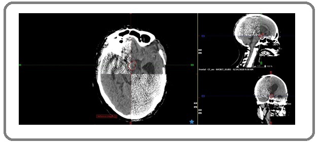
The treatment plan was evaluated by radiation oncologist and medical physicist against the single fraction RTOG organs at risk (OARs). Before delivering dose to patient, patient specific quality assurance (QA) was done to assure prescribed dose would be delivered accurately within acceptable limits. Patient specific QA included collision check, dummy run and dosimetric fluence variation. The plan was delivered to phantom. The measured fluence was evaluated against TPS calculated fluence. QA tests were passed with 100% scores. The patient was positioned on treatment couch; stereotactic frame (DSPS) was fitted on patient’s head. Isocenters were matched and shifts were applied according to plan made. Reproducibility was assessed KVCT to ensure ±1 mm accuracy and precision of treatment (Figure 3).
Figure 3. Showing the co-axial Registration of Onboard kVCT Obtained Just before Treatment with Planning CT.

Treatment was delivered by Varian True Beam with 6 FFF.
Outcome
On his first follow up at 1-month post SRS, he was completely free of symptoms. MRI Brain was done 6 months’ post SRS which was suggestive of reduction in size of lesion (10 mm × 10 mm Vs 24 mm × 14 mm).
6 months’ post treatment; he is completely free of symptoms.
Discussion
Craniopharyngioma is a low grade tumour but its close proximity to organs like pituitary, hypothalamus, brainstem and optic chiasm causes significant endocrinological, neurological and visual sequelae [2-4].
Subtotal resection (STR) is preferred over gross total resection due to anatomical proximity to hypothalamus. However, STR is associated with high rates of recurrence. So, adjuvant EBRT is added to reduce the recurrence [6]. Our patient did not receive adjuvant radiotherapy and instead kept on observation. He developed recurrence 1 year after STR. This could have been avoided with use of adjuvant RT.
When he developed symptoms due to recurrence of lesion, he presented to our hospital for further evaluation and treatment. He was evaluated by multi-disciplinary team (neurosurgery, neuro radiology and radiation oncology). Repeat debulking was not feasible for him so, we proceeded with RT. RT could be delivered with either conventional fractionation or advanced techniques like SRT or SRS. Studies have shown excellent result with SRS for small tumour volume and tumours > 3mm away from critical structures like optic chiasm, nerve and brainstem [7, 8]. As our patient tumour characteristic fulfilled the criteria for SRS treatment, we proceeded with same.
Here, we reviewed a patient of craniopharyngioma who underwent STR and developed recurrence. He was treated with SRS as second debulking surgery was not feasible. He demonstrated an excellent response following completion of treatment.
References
- Primer'Craniopharyngioma'. NATURE REVIEWS DISEASE PRIMERS Mueller HL, Merchant TE, Warmuth-Metz M, Martinez-Barbera JP, Puget S. 2019 .2019 Nov 7;5.
- Craniopharyngiomas Karavitaki Niki, Cudlip Simon, Adams Christopher B. T., Wass John A. H.. Endocrine Reviews.2006;27(4). CrossRef
- Aggressive surgical management of craniopharyngiomas in children Hoffman Harold J., Silva Marcia De, Humphreys Robin P., Drake James M., Smith Mary Lou, Blaser Susan I.. Journal of Neurosurgery.1992;76(1). CrossRef
- Craniopharyngioma. Medical Care Bobustuc GC, Groves MD, Fuller GN, DeMonte F. 28.2002;:58-78.
- Bernal-Sprekelsen M, Braun H, Cappabianca P, Carrau R. European position paper on endoscopic management of tumours of the nose, paranasal sinuses and skull base Lund VJ, Stammberger H, Nicolai P, Castelnuovo P, Beal T, Beham A. Rhinol Suppl.2010;22(2):1-43.
- The role of radiotherapy in the treatment of craniopharyngeoma-indications, results, side effects Becker G, Kortmann RD, Skalej M, Bamberg M. Frontiers of radiation therapy and oncology.1999;33(33):100-113. CrossRef
- Long-term results of gamma knife surgery for the treatment of craniopharyngioma in 98 consecutive cases Kobayashi Tatsuya, Kida Yoshihisa, Mori Yoshimasa, Hasegawa Toshinori. Journal of Neurosurgery: Pediatrics.2005;103(6). CrossRef
- Stereotactic radiosurgery of residual or recurrent craniopharyngioma, after surgery, with or without radiation therapy Chiou Shang-Ming, Lunsford L. Dade, Niranjan Ajay, Kondziolka Douglas, Flickinger John C.. Neuro-Oncology.2001;3(3). CrossRef
License

This work is licensed under a Creative Commons Attribution-NonCommercial 4.0 International License.
Copyright
© Asian Pacific Journal of Cancer Care , 2021
Author Details
How to Cite
- Abstract viewed - 0 times
- PDF (FULL TEXT) downloaded - 0 times
- XML downloaded - 0 times