Capacity of Thymidylate Synthase and Dihydropyrimidine Dehydrogenase mRNA Expression to Predict Neoadjuvant Chemotherapy Response of 5fu Regimen in Advanced Colorectal Cancer: A Cross-Sectional Study
Download
Abstract
Background: Chemotherapy is an important component of colorectal cancer treatment. Despite its success in inducing tumor cell death, chemotherapy has been constrained by resistance and adverse side effects. Therefore, predictive markers are required to ensure that the appropriate chemotherapy is administered based on the individual patient. This study aimed to correlate the mRNA expression of the TS and DPD genes with the tumor and CEA responses to the 5FU chemotherapy regimen.
Methods: This was a cross-sectional study with a prognostic test design. Measurements of tumor mass and tissue sampling were done prior to chemotherapy. CT scans were performed to measure tumor size pre-chemotherapy and after two cycles of treatment with a lag time of three weeks. The mRNA expression of the TS and DPD genes was then evaluated in each group.
Results: The mean value of the mRNA expression of the TS gene (9.555±1.693) showed an insignificant correlation with clinical response p=0.195 (p<0.05). However, the mean value of the mRNA expression of the DPD gene (7.461±1.088) was significantly correlated with clinical response p=0.003 (p<0.05).
Conclusion: DPD mRNA can be considered a marker of chemotherapy responsiveness when choosing a chemotherapy regimen.
Introduction
Colorectal cancer (CRC) is the fourth most common malignancy in men and the third most common in women worldwide [1]. CRC has a global incidence of 1 million new cases and a mortality of half a million people annually. In Indonesia, the incidence of colorectal cancer incidence in 2020 is 12.8 cases per 100,000 population, and it is increasing each year [2]. Local recurrence after treatment is reported in 3–32 % of patients [3, 4].
Chemotherapy is an important component of colorectal cancer therapy. Although it has proven successful in inducing tumor cell death, chemotherapy has been constrained by chemo-resistance and adverse side effects. For over five decades, 5-fluorouracil (5-FU) has been incorporated into many standard CRC chemotherapy treatments [3]. Adverse reactions to 5-FU-based chemotherapy have been reported to be caused by genetic variants of the drug-related thymidylate synthase (TS; TYMS) and dihydropyrimidine dehydrogenase (DPD; DPYD) genes. Thymidylate synthase is an important target for chemotherapeutic drugs such as 5-FU and methotrexate; however, the overexpression of TS can cause resistance to targeted treatments. Dihydropyrimidine dehydrogenase (DPD) is the catabolic enzyme of 5-FU and is related to the chemotherapy responsiveness to 5-FU [5, 6]. Therefore, we conducted this study to investigate the correlation between the expression of the mRNA of the TS and DPD genes and the tumor and CEA responses to the 5-FU chemotherapy regimen.
Materials and Methods
This research involved a cross-sectional study at the surgery clinic of Bahtera Mas Hospital, Kendari, Southeast Sulawesi, Indonesia covering the period between January 2023 and October 2023. The Ethics Committee of the Faculty of Medicine at the University of Hasanuddin approved the research protocols (number: 90/UN4.6.4.5.31/PP36/2024) on February 20, 2024. This study was registered with the Thai Research Registry (TCTR20240618004) and was conducted in accordance with The Code of Ethics of the World Medical Association (Declaration of Helsinki). Informed consent was obtained from all participants involved in the study.
The sample size was determined using the formula designed for testing the hypothesis of the difference in means of two independent populations, using a minimum of 30 participants [7, 8]. The inclusion criteria consisted of patients with Stage III or IV colorectal cancer, histopathology results obtained through biopsy showing adenocarcinoma, and a willingness to undergo adjuvant chemotherapy with a regimen of capecitabine and oxaliplatin. The exclusion criteria were damaged tissue samples, patients who refused chemotherapy follow-up, and pathology results showing signet ring cell or mucinous carcinoma.
Research procedure
We performed biomolecular examinations of patient samples pre- and post-chemotherapy. Tumor mass and tissue measurements were conducted prior to chemotherapy, and CT scans were performed to measure tumor size pre-chemotherapy and after two cycles of chemotherapy with a lag time of three weeks. The measurements were repeated after four cycles of chemotherapy.
The clinical responses of the patients were measured based on the tumor size differences pre- and post- chemotherapy. The percentage change was interpreted based on the response evaluation criteria in solid tumor (RECIST 1.1) criteria. A non-responsive result was determined when the disease was considered stable or progressive (tumor size decreased <30 %), tumor size was maintained or increased, or new tumors were found. A responsive result was defined as a partial or complete response if the tumor mass disappeared, the tumor size decreased by ≥30%, and no new tumors were found.
The Bahtera Mas Hospital samples were sent to the Microbiology Laboratory at the Faculty of Medicine, University of Hasanuddin, Makassar, South Sulawesi for qRT-PCR examination. Tissue samples were immediately placed into container bottles containing “L6” solution (consisting of 120 g of guanidine thiocyanate (GuSCN) in 100 mL 0.1 M Tris HCl pH 6.4, 22 mL 0.2 M.
Ethylenediamine Tetra-Acetate (EDTA) pH 8.0, and 2.6 g Triton X-100 to a final concentration of 50 mM Tris HCl, 5 M GuSCN, 20 mM EDTA, 0.1 % Triton X-100) [9].
The qRT-PCR Procedure (Boom Method)
We used a quantitative real-time PCR system (Applied Biosystems, Waltham, MA, USA) using Power SYBR Green. The 50 μL reaction mix containing 25 μL Premix Ex Taq (2×, TaKaRa), 1 μL ROX reference Dye II (50×, TaKaRa), 1 μL PCR forward primer (10 μM), 1 μL PCR reverse primer (10 μM), 4 μL cDNA, and 18 μL dH2O was transferred to 96-well plates. The primary sequences to detect TS mRNA were the reverse primer, Thymidylate Synthase: forward, 5′-GCC AGA ATC TGT TCG CTT CAA C-3′ , reverse, 5′-AGG AAA CTG AGT GCC GGC TT-3′; and the reference gene(β-actin gene): forward, 5′-CCT CCA TCA TCC TCT GTT CTA CTC T-3′, and reverse, 5′-TGC TCT CAT ATG CAG AAG CTA GAA A-3′. The primary sequences to detect DPD were the reverse primer: 5ʹ-CTTTGGGTGCGACTTGACG-3ʹ and 5ʹ-GTCGACCCCGCTCCTTTT-3ʹ; and GAPDH, primers sequences: 5ʹ-AACAGCGACACCCACTCCTC- 3ʹ and 5ʹ-GGAGGGGAGATTCAGTGTGGT-3ʹ. [9] We performed the analysis in triplicate and the standard curve indicated good efficiency of amplification (90–100 %). We then determined the cut-off for each gene expression level using an ROC curve analysis.
Statistical Analysis
The statistical analysis was conducted using the Statistical Package for the Social Sciences (SPSS). The Wilcoxon signed test, Mann-Whitney U test and Spearman rho test were used. A p-value <0.05 was considered significant. After obtaining the results of the qRT-PCR examination of the TS and DPD genes, the cut-off was determined using an ROC analysis and the AUC.
Results
Patient Characteristics
Forty-two patients met the study criteria and were sampled; however, 6 withdrew, leaving 36 participants for the analysis (Table 1).
| n (%) | Mean (SD) | ||
| Variable | Category | ||
| Sex | Men | 24 (66.7) | |
| Women | 12 (33.3) | ||
| Age (years) | ≤50 | 25 (69.4) | 41.52 (5.38) |
| >50 | 11 (30.6) | 55.00 (41.23) | |
| Tumor size (cm) | Pre-chemotherapy | 36 | 13.28 (4.77) |
| Post-chemotherapy | 36 | 9.28 (4.74) | |
| Chemotherapy response | Responsive | 18 (50.0) | |
| Non-responsive | 18 (50.0) | ||
| Low | 16 (44.4) | ||
| Grading | Moderate | 17 (47.3) | |
| High | 3 (8.3) |
Of these, 24 (66.7 %) were male and 25 (69.4 %) were ≤50 years old; the mean age was 46 years. Seventeen participants (47.2 %) had tumors in the rectum, 7 (19.4 %) in the ascending colon, 7 (19.4%) in the sigmoid colon, 3 (8.3 %) in the transverse colon, and 2 (5.6 %) had tumors in the descending colon. Most of the patients (44.4 %) had low-grade cancer. The CEA response, which is a biomarker indicator, revealed that 14 people (38.9 %) had good responses to the treatment. However, based on tumor size, only 18 patients (50 %) were chemotherapy responsive. The statistical analysis showed a significant association between sex (p = 0.014), pathological result (p = 0.046), CEA response (p = 0.048), tumor response (p = 0.007), and clinical response (p < 0.05).
Tumor Size and CEA with Chemotherapy Response
The statistical analysis showed a significant decrease in tumor size and CEA level after the administration of chemotherapy compared to those values before treatment (p<0.05) (Table 2).
| Variable | Pre-chemotherapy | Post-chemotherapy | p-value* |
| (Mean ± SD) | (Mean ± SD) | ||
| Tumor size (cm) | 9.53 ± 3.80 | 6.50 ± 3.30 | <0.001 |
| CEA (ng/mL) | 8.67 ± 6.02 | 6.30 ± 5.70 | <0.001 |
Note, *Wilcoxon test
In addition, we analyzed the mean differences in tumor size reduction and CEA decrease between the responsive and non-responsive groups (Table 3).
| Variable | Chemotherapy Response | p-value* | |
| Responsive | Non-Responsive | ||
| (Mean ± SD) | (Mean ± SD) | ||
| Δ Tumor Size (cm) | 2.14 ± 1.56 | 2.52 ± 2.12 | 0.81 |
| Δ CEA (ng/mL) | 1.36 ± 1.74 | 4.09 ± 2.65 | <0.001 |
Note, Mann-Whitney U test
The difference in biomarkers based on chemotherapy response in colorectal carcinoma.
The Mann-Whitney U test revealed that the mRNA levels of the TS gene did not differ significantly (p>0.05); therefore, the mRNA of the TS gene was not able to distinguish patients based on their responsiveness to chemotherapy (Table 4).
| Biomarker | Responsive | Non-Responsive | p-value* | ||
| Median | Min/Max | Median | Min/Max | ||
| TS mRNA gene | 9 | 7.5/13.1 | 8.95 | 8.3/13.5 | 0.584 |
| DPD mRNA gene | 7.73 | 4.5/11.2 | 8.13 | 4.7/11.5 | 0.037 |
*Mann-Whitney U test
However, the mRNA levels of the DPD gene differed significantly (p<0.05), whereby the mRNA levels of the DPD gene were lower in the responsive patients than those in the non-responsive participants (median 7.73 vs 8.13, respectively).
The Influence of mRNA Expression of the TS and DPD Genes on Tumor Size Change
The correlation analysis (Table 5), based on the Spearman correlation coefficient (r) and p-value, revealed no significant correlation between mRNA expression of the TS gene and change in tumor size (r=-0.143, p=0.406).
| Variable | Correlation coefficient (r) | p-value |
| TS gene mRNA | -0.143 | 0.406 |
| DPD gene mRNA | -0.296 | 0.08 |
| Combined mRNA of TS and DPD genes | -0.163 | 0.342 |
Note; r=Spearman
Similarly, there was no significant relationship between mRNA expression of the DPD gene and change in tumor size (r=-0.296, p=0.080).
Furthermore, the combination of the two genes returned similar results, whereby the correlation with tumor size was not significant (r=-0.163, p=0.342). Therefore, mRNA expression of the TS and DPD genes had no influence on tumor size change in this study.
mRNA Expression of TS Gene and Chemotherapy Response
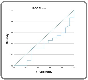
The ROC analysis showed a cut-off of TS mRNA expression at 8.66 with a sensitivity of 72.7 %, a specificity of 92.9 %, and an accuracy of 55.6 % (Figure 1).
Figure 1. ROC Analysis of TS Gene mRNA as a Predictor of Tumor Size.

The mean value of mRNA expression of the TS gene was 9.555±1.693. In the chemotherapy-responsive group, the median value of mRNA expression of the TS gene was 9 (7.5–13.1), whereas the median value was 8.95 (8.3–13.5) in the non-responsive group. The mRNA expression of the TS gene was not significantly associated with clinical response (p=0.195). Similarly, no significant correlation was found between the TS gene mRNA and tumor size change (r=-0.143, p=0.406).
mRNA Expression of DPD Gene and Chemotherapy Response
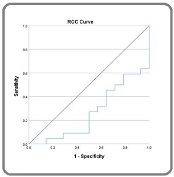
After obtaining the results of the qRT-PCR examination of the DPD gene, the cut-off was determined using an ROC analysis and the AUC. The cut-off of DPD mRNA expression was 7.59 with a sensitivity of 63.6 %, a specificity of 92.9 %, and an accuracy of 70.4 % (Figure 2).
Figure 2. ROC Analysis of DPD Gene mRNA as a Predictor of Tumor Size.

The mean value of mRNA expression of the DPD gene was 7.461±1.088. The chemotherapy-responsive and non-responsive groups showed median mRNA expression values of the DPD gene of 7.73 (4.5–11.2) and 8.13 (4.7–11.5), respectively. The DPD gene mRNA expression was significantly associated with clinical response (p=0.003), although no significant correlation was found between DPD gene mRNA and tumor size change (r=-0.296, p=0.080).
Discussion
The results of this study showed a significant association between sex (p=0.014), pathological result (p=0.046), CEA response (p=0.048), tumor response (p=0.007), and clinical response (p<0.05). Age is a major risk factor for CRC, and this was confirmed in this study, as the number of CRC cases increased in individuals over the age of 40 years. Previous studies have found that CRC cases rarely occur before the age of 40, begin to increase between the ages of 40 and 50, and decrease in older groups [10-17]. The increase in the incidence of CRC is highly affected by screening, and since many cancers are first detected by screening at the age of 50 years, CRC screening should begin at this age [12]. Regarding stage stratification, many cases of invasive cancer are identified in patients over 49 years of age due to late diagnosis [16]. The morbidity and mortality of colorectal cancer in individuals over 49 are increasing, in which 29 % to 30 % have a 5-year mortality rate and require surgery and chemotherapy. Sanoff et al. found that the response to 5FU-Oxaliplatin chemotherapy was dependent on age, with increased survival in patients under 75 years [18].
This study found that male sex was associated with chemotherapy response (p=0.014). Previous studies have shown a lower survival rate in women than men [19], which is possibly a result of genetic and epigenetic differences in the sexes. Women show a higher percentage of developing tumors in the cecum from high levels of CIMP from the rectum to the cecum [20], and PIK3CA mutations are more common in women, which is associated with poor survival [21]. CRC screening results in women often find larger polyp sizes than those in men, and clinical treatment studies show that a high percentage of women experience recurrence after 5FU-oxaliplatin chemotherapy and persistent amenorrhea for one year after treatment, which can affect menopause and fertility [22].
In this study, tumor location was not associated with chemotherapy response. However, previous studies have found that tumor location can be a prognostic factor in CRC patient survival. The CRC location most commonly found in this study was the rectum. Patients with CRC in the rectum have shorter survival potentials than those with CRC in the cecum, transverse colon, descending colon, sigmoid colon, and rectosigmoid regions [23]. Survival based on tumor location is related to the location of metastases [24,25]. Based on biological factors, the survival of patients with rectal cancer corresponds to the increasing number of mutations in BRAF and KRAS and is associated with a poor prognosis [26]. However, Bozkurt et al. and Ryuk et al. determined that tumor location was not a risk factor for CRC [27,28]. The most common locations for CRC are the ascending and sigmoid colons. These regions are often exposed to feces, causing mucosal changes in the colon that eventually become tumors [23]. In this study, most participants were responsive to 5FU-oxaliplatin, which can exert antitumor activity by inducing thymidylate deficiency and balancing the nucleotide pool, thereby disrupting DNA replication, transcription, and repair and causing cell death [29]. In addition, administering combination chemotherapy treatments can result in the more efficient destruction of CRC tumor cells [30]. However, resistance to 5-FU chemotherapy and oxaliplatin has been found in patients with colorectal cancer [31, 32].
This study used the chemotherapy combination of 5FU and oxaliplatin; however, the use of oxaliplatin is known to cause resistance. This resistance is caused by an increase in the ATP-binding cassette drug transporter, which reduces the intracellular concentration of oxaliplatin. In addition, an increase in oxaliplatin protein (mammalian metallothionein-2A) MT2A [33], which is a protein that can inhibit apoptosis and increase proliferation in CRC cells can cause resistance [34]. However, resistance to the combination of 5-FU/LV and oxaliplatin has not been identified; therefore, this study is novel in the selection of chemotherapy treatments for advanced CRC. Understanding the mechanism of chemoresistance is crucial in the development of new and more effective treatment combinations.
mRNA Expression of TS Gene and Chemotherapy Response
Our study found the mean value of mRNA expression of the TS gene (9.555±1.693), had an insignificant correlation with clinical response p=0.195 (p<0.05). Shirota et al. found that the level of TS mRNA expression can be used as a tumor marker for CRC patients undergoing 5FU chemotherapy [35], whereas Kuramochi et al. stated that patients with high TS mRNA expression showed lower colorectal tumor responses [36]. The TS inhibitor 5-fluorouracil (5-FU) is widely used for the treatment of colorectal, pancreatic, gastric, and ovarian cancers, and leucovorin, which is a folate-reducing agent, has been shown to increase the activity of 5-FU in colorectal cancers. However, response rates for chemotherapy combinations are approximately 25 %–30 % and much effort has been focused on designing new, more potent TS inhibitors, such as capecitabine, which has been approved by the US Food and Drug Administration as first-line therapy for patients with advanced colorectal cancer. In addition, TS protein and TS mRNA expression are highly correlated and able to predict the response to 5-FU/LV-based chemotherapy in patients with colorectal and gastric cancers, respectively. Lower TS gene expression levels correspond to higher tumor responses in CRC patients undergoing 5FU chemotherapy [37].
mRNA Expression of DPD Gene and Chemotherapy Response
The mean mRNA expression value of the DPD gene in this study (7.461±1.088) was significantly correlated with clinical response p=0.003 (p<0.05). Similar to the results for TS, Shirota et al. determined that DPD mRNA expression level was an effective tumor marker for CRC patients undergoing 5FU chemotherapy [35]. However, Kuramochi et al. reported no significant relationship between patients with DPD mRNA expression and chemotherapy response [36].
Uchida et al. found that DPD was a key enzyme in the catabolic pathway of 5-FU. Significant differences were noted in DPD mRNA levels in colorectal cancers during chemotherapy. The results of this study suggest that 5FU may affect DPD mRNA expression in colorectal cancer patients, while TS/DPD expression may be considered an independent prognostic factor and colorectal cancer patients with low DPD mRNA expression could benefit from 5FU-based neoadjuvant chemotherapy. In addition, quantitative analysis of DPD mRNA changes in surgical specimens during 5FU-based chemotherapy may predict the disease-free interval for postoperative colorectal cancer patients more effectively than endoscopic specimen analysis before chemotherapy. Furthermore, TS and DPD regulation appear to be associated during 5FU chemotherapy. Elucidation of the mechanisms that regulate TS and DPD mRNA expression may allow for the prediction of sensitivity and/or toxicity to 5FU [38]. Lower DPD gene expression values equate to higher tumor responses in CRC patients who undergo 5FU chemotherapy [37]. Al-Rubaiawi et al. showed that serum and tissue DPD activities in advanced-stage CRC patients were high when compared to those of early-stage CRC patients and the control. In addition, early-stage CRC patients returned higher DPD activities than those of the controls. The DPD levels in tissues of advanced and early CRC patients were significantly different from those in normal tissues, although no significant differences were found in mean serum DPD levels between CRC patients (early and advanced) and healthy controls. In addition, DPD showed a moderate correlation with CA19-9 in CRC patients (early and advanced) that approached significance [39].
One limitation of this study was the level of patient dropout, although the remaining number of participants allowed for the minimum sample size to be met. In addition, we only included patients at our center; therefore, we cannot describe the same conditions in different populations and locations. However, a strength of this study was the cross-sectional observational research and prognosis test design, which is effective for determining the usefulness of variables in predicting certain future outcomes. In addition, this study analyzed clinicopathological factors that theoretically affect chemotherapy response, thereby allowing the results obtained to confirm the relationships between variables. In conclusion, the mRNA expression of the TS gene had no effect on the clinical response to 5FU neoadjuvant chemotherapy in advanced colorectal cancer, whereas the mRNA expression of DPD gene influenced this clinical response. A cut-off value for the mRNA expression of the DPD gene of ≤7.9 would produce a good response to the chemotherapy. Therefore, we suggest that mRNA expression of the DPD gene can be used as a marker of chemotherapy responsiveness and should be considered when choosing a chemotherapy regimen.
Acknowledgements
None
Author contributions
Conception: ECT, AAI, MH, RL, PRI, RB, IKW, IJP,
and AB; Interpretation or analysis of data: ECT, AAI, MH, RL, PRI, RB, and IKW; Preparation of the manuscript: ECT, AAI, MH, RL, PRI, and MF; Revision for important intellectual content: ECT, AAI, MH, RL, PRI, RB, and IKW; Supervision: AAI, MH, RL, PRI, RB, and IKW. All authors read and approved the final version of the manuscript.
Conflict of Interest
The authors declare that they have no conflict of interest.
Funding
Self-funding
References
- Cancer statistics, 2013 Siegel R, Naishadham D, Jemal A. CA: a cancer journal for clinicians.2013;63(1). CrossRef
- Global Cancer Statistics 2020: GLOBOCAN Estimates of Incidence and Mortality Worldwide for 36 Cancers in 185 Countries Sung H, Ferlay J, Siegel RL , Laversanne M, Soerjomataram I, Jemal A, Bray F. CA: a cancer journal for clinicians.2021;71(3). CrossRef
- Kementerian Kesehatan Republik Indonesia. Panduan Penatalaksanaan Kanker Kolorektal. Jakarta: Kemenkes RI 2014.
- Cancer Incidence and Mortality in a Tertiary Hospital in Indonesia: An 18-Year Data Review Prihantono n, Rusli R, Christeven R, Faruk M. Ethiopian Journal of Health Sciences.2023;33(3). CrossRef
- Thymidylate synthase gene polymorphism predicts toxicity in colorectal cancer patients receiving 5-fluorouracil-based chemotherapy Lecomte T, Ferraz J, Zinzindohoué F, Loriot M, Tregouet D, Landi B, Berger A, et al . Clinical Cancer Research: An Official Journal of the American Association for Cancer Research.2004;10(17). CrossRef
- DPD Testing Before Treatment With Fluoropyrimidines in the Amsterdam UMCs: An Evaluation of Current Pharmacogenetic Practice Martens FK , Huntjens DW , Rigter T, Bartels M, Bet PM , Cornel MC . Frontiers in Pharmacology.2019;10. CrossRef
- Memon MA, Ting H, Cheah J-H, Thurasamy R, Chuah F, Cham TH. Sample Size for Survey Research: Review and Recommendations. Journal of Applied Structural Equation Modeling 2020;4:i–xx. doi: 10.47263/JASEM.4(2)01. .
- Research Methods For Business: A Skill Building Approach, 7th ed. London: John Wiley & Sons, Ltd Sekaran U, Bougie R, Sampling. In: Sekaran U, Bougie R, eds . 2016;:448.
- Rapid and simple method for purification of nucleic acids Boom R., Sol C. J., Salimans M. M., Jansen C. L., Wertheim-van Dillen P. M., Noordaa J.. Journal of Clinical Microbiology.1990;28(3). CrossRef
- Increasing disparities in the age-related incidences of colon and rectal cancers in the United States, 1975-2010 Bailey CE , Hu C, You YN , Bednarski BK , Rodriguez-Bigas MA , Skibber JN , Cantor SB , Chang GJ . JAMA surgery.2015;150(1). CrossRef
- National Trends in Colorectal Cancer Incidence Among Older and Younger Adults in Canada Brenner DR , Heer E, Sutherland RL , Ruan Y, Tinmouth J, Heitman SJ , Hilsden RJ . JAMA network open.2019;2(7). CrossRef
- Trends in Incidence of Early-Onset Colorectal Cancer in the United States Among Those Approaching Screening Age Abualkhair WH , Zhou M, Ahnen D, Yu Q, Wu X, Karlitz JH . JAMA network open.2020;3(1). CrossRef
- Trends in the Incidence of Young-Onset Colorectal Cancer With a Focus on Years Approaching Screening Age: A Population-Based Longitudinal Study Howren A, Sayre EC , Loree JM , Gill S, Brown CJ , Raval MJ , Farooq A, De Vera MA . Journal of the National Cancer Institute.2021;113(7). CrossRef
- Trends in Incidence and Stage at Diagnosis of Colorectal Cancer in Adults Aged 40 Through 49 Years, 1975-2015 Meester RGS , Mannalithara A, Lansdorp-Vogelaar I, Ladabaum U. JAMA.2019;321(19). CrossRef
- Contributions of Adenocarcinoma and Carcinoid Tumors to Early-Onset Colorectal Cancer Incidence Rates in the United States Montminy EM , Zhou M, Maniscalco L, Abualkhair W, Kim MK , Siegel RL , Wu , et al . Annals of Internal Medicine.2021;174(2). CrossRef
- Colorectal Cancer Initial Diagnosis: Screening Colonoscopy, Diagnostic Colonoscopy, or Emergent Surgery, and Tumor Stage and Size at Initial Presentation Moreno CC , Mittal PK , Sullivan PS , Rutherford R, Staley CA , Cardona K, Hawk NN , Dixon WT , et al . Clinical Colorectal Cancer.2016;15(1). CrossRef
- Annual Report to the Nation on the Status of Cancer, Featuring Cancer in Men and Women Age 20-49 Years Ward EM , Sherman RL , Henley SJ , Jemal A, Siegel DA , Feuer EJ , Firth AU , et al . Journal of the National Cancer Institute.2019;111(12). CrossRef
- Effect of adjuvant chemotherapy on survival of patients with stage III colon cancer diagnosed after age 75 years Sanoff HK , Carpenter WR , Stürmer T, Goldberg RM , Martin CF , Fine JP , McCleary NJ , et al . Journal of Clinical Oncology: Official Journal of the American Society of Clinical Oncology.2012;30(21). CrossRef
- Data on the characteristics and the survival of korean patients with colorectal cancer from the Korea central cancer registry Park H, Shin A, Kim B, Jung K, Won Y, Oh J, Jeong S, et al . Annals of Coloproctology.2013;29(4). CrossRef
- Prognostic implication of the CpG island methylator phenotype in colorectal cancers depends on tumour location Bae J. M., Kim J. H., Cho N.-Y., Kim T.-Y., Kang G. H.. British Journal of Cancer.2013;109(4). CrossRef
- Descriptive profile of PIK3CA-mutated colorectal cancer in postmenopausal women Phipps AI , Makar KW , Newcomb PA . International Journal of Colorectal Disease.2013;28(12). CrossRef
- Incidence of chemotherapy-induced amenorrhea in premenopausal women treated with adjuvant FOLFOX for colorectal cancer Cercek A, Siegel CL , Capanu M, Reidy-Lagunes D, Saltz LB . Clinical Colorectal Cancer.2013;12(3). CrossRef
- Primary tumor location as a prognostic factor in metastatic colorectal cancer Loupakis F, Yang D, Yau L, Feng S, Cremolini C, Zhang W, Maus MKH , et al . Journal of the National Cancer Institute.2015;107(3). CrossRef
- Patterns of metastasis in colon and rectal cancer Riihimäki M, Hemminki A, Sundquist J, Hemminki K. Scientific Reports.2016;6. CrossRef
- Stage IV colorectal cancer primary site and patterns of distant metastasis Robinson JR , Newcomb PA , Hardikar S, Cohen SA , Phipps AI . Cancer Epidemiology.2017;48. CrossRef
- Clinicopathological characteristics and prognostic impact of colorectal cancers with NRAS mutations Ogura T, Kakuta M, Yatsuoka T, Nishimura Y, Sakamoto H, Yamaguchi K, Tanabe M, Tanaka Y, Akagi K. Oncology Reports.2014;32(1). CrossRef
- Conservative treatment of scapular neck fracture: the effect of stability and glenopolar angle on clinical outcome Bozkurt M, Can F, Kirdemir V, Erden Z, Demirkale I, Başbozkurt M. Injury.2005;36(10). CrossRef
- Predictive factors and the prognosis of recurrence of colorectal cancer within 2 years after curative resection Ryuk JP , Choi GS , Park JS , Kim HJ , Park SY , Yoon GS , Jun SH , Kwon YC . Annals of Surgical Treatment and Research.2014;86(3). CrossRef
- ABCB5 identifies a therapy-refractory tumor cell population in colorectal cancer patients Wilson BJ , Schatton T, Zhan Q, Gasser M, Ma J, Saab KR , Schanche R, et al . Cancer Research.2011;71(15). CrossRef
- Pharmacologic resistance in colorectal cancer: a review Hammond WA , Swaika A, Mody K. Therapeutic Advances in Medical Oncology.2016;8(1). CrossRef
- Anemoside B4 sensitizes human colorectal cancer to fluorouracil-based chemotherapy through src-mediated cell apoptosis He X, Tang J, Yan H, Wang J, Li H, Duan X, Yu S, Hou X, Liao G, Liu W. Aging.2021;13(23). CrossRef
- Long noncoding RNA CRART16 confers 5-FU resistance in colorectal cancer cells by sponging miR-193b-5p Wang J, Zhang X, Zhang J, Chen S, Zhu J, Wang X. Cancer Cell International.2021;21(1). CrossRef
- MT2A Promotes Oxaliplatin Resistance in Colorectal Cancer Cells Zhao Z, Zhang G, Li W. Cell Biochemistry and Biophysics.2020;78(4). CrossRef
- Metallothionein 2A an interactive protein linking phosphorylated FADD to NF-κB pathway leads to colorectal cancer formation Marikar FMMT , Jin G, Sheng W, Ma D, Hua Z. Chinese Clinical Oncology.2016;5(6). CrossRef
- ERCC1 and thymidylate synthase mRNA levels predict survival for colorectal cancer patients receiving combination oxaliplatin and fluorouracil chemotherapy Shirota Y., Stoehlmacher J., Brabender J., Xiong Y. P., Uetake H., Danenberg K. D., Groshen S., et al . Journal of Clinical Oncology: Official Journal of the American Society of Clinical Oncology.2001;19(23). CrossRef
- 5-fluorouracil-related gene expression levels in primary colorectal cancer and corresponding liver metastasis Kuramochi H, Hayashi K, Uchida K, Miyakura S, Shimizu D, Vallbohmer D, Park S, et al . International Journal of Cancer.2006;119(3). CrossRef
- Tumor 5-FU-related mRNA Expression and Efficacy of Oral Fluoropyrimidines in Adjuvant Chemotherapy of Colorectal Cancer Koda K, Miyauchi H, Kosugi C, Kaiho T, Takiguchi N, Kobayashi S, Maruyama T, Matsubara H. Anticancer Research.2016;36(10). CrossRef
- Changes in intratumoral thymidylate synthase (TS) and dihydropyrimidine dehydrogenase (DPD) mRNA expression in colorectal and gastric cancer during continuous tegafur infusion Uchida K., Hayashi K., Kuramochi H., Takasaki K.. International Journal of Oncology.2001;19(2). CrossRef
- The Activity of 5-Flurouracil Metabolizing Enzyme Dihydropyrimidine Dehydrogenase(DPD) and its Association with Tumor Progression and Markers (CEA, CA19.9) in Patients with Colorectal Cancer AL-Rubaiawi HK , Mohamed RJ . Indian J Public Health Res Dev.2019;10:2363. CrossRef
License

This work is licensed under a Creative Commons Attribution-NonCommercial 4.0 International License.
Copyright
© Asian Pacific Journal of Cancer Biology , 2025
Author Details